Rajasthan Board RBSE Class 12 Biology Notes Chapter 24 Man-Blood Vascular, System
General
The circulatory system is required to transport nutrients to various body organs and to remove waste products. Unicellular or monoblastic/diploblastic multicellular organisms take 02 and nutrients directly from environment and release C02 and wastes directly to environment.
In multi cellular organisms cells are not in direct contact with the environment so in there is a need of a system which exchanges the gases, circulate the food materials and release waste material into the environment. This system is known as circulatory system.
It is of two types-
- Open Circulatory system:
In open circulatory system liquid flows between cells and tissues, openly i.e. does not flow in closed blood vessels.
Example – Molluses and Arthropods. - Closed Circulatory system:
In closed circulatory system, the fluid flows within closed vessels.
Example – Human and Rabbit etc.
![]()
Blood
General
- It is a red coloured liquid connective tissue which originates from the mesoderm.
- It reaches into the various organs through the blood vessels and transports various chemical substances to different tissues.
- The specific gravity of blood is 1.04 & it’s viscosity is 3. times more than the water.
- The blood it salty in taste & alkaline (pH-7.4) in nature.
- The amount of blood is 8% by weight. Hence, a person of 70 kg weight has about 5.6 litre of blood. Study of blood is called as haematology.
- The blood has two parts viz. plasma & blood corpuscles.
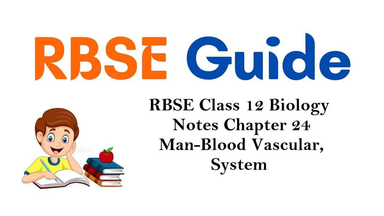
Plasma
- It is light yellow slightly alkaline liquid and it is 55% of blood. It is made up of 90% water and 10% inorganic and organic substances which are found in soluble or colloidal state.
- Proteins are the most abundantly found organic substance in Plasma (7%) and inorganic substances are 0.09% out of which Sodium ion is the most.
- Main components of plasma and their functions are listed in Table
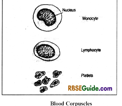
Blood Corpuscles
They are 45% of the total blood and are of three types-
- Red Blood corpuscles [RBC]
- White Blood corpuscles [WBC]
- Platelets or Thrombocytes
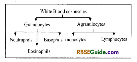
RBC or Erythrocvtes
They are the main blood corpuscles.
Each RBC is small, round, disc-like, biconcave & enucleated. It’s deameter is about 7.5ft & thickness is 1 to 2 μ.
The life span of RBC is of 50 to 120 days.
RBCs are formed in red bone marrow from myeloblasts & the process of formation is called as erythropoeisis. This process takes about 72 hours.
During embryonic stage, RBC are formed mainly in liver but also in the ribs & spleen.
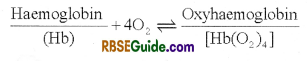
RBC is full of haemoglobin which acts as a respiratory pigment.
The amount of Hb in man is 14-16 gm% & in woman is 12-14 gm%.
Dead RBCs from the blood is removed in the liver & spleen. The liver breaks Hb of dead RBC & form bilirubin (yellow) & biliverdin (green) as bile pigments. They are excreted along with the bile juice.
The group of plasmalemma of dead RBCs is called a ghost of RBC which is destroyed in the spleen.
One molecule of haemoglobin transports four molecules [4O2] of oxygen as oxyhemoglobin.
![]()
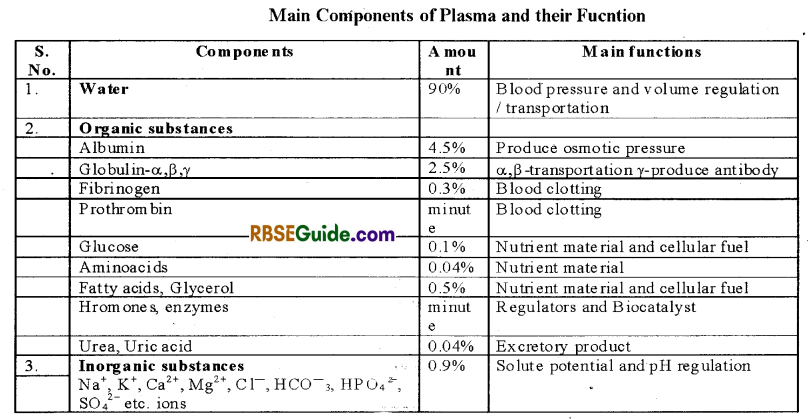
WBC or Leucocytes
They are nucleated, colourless & complete cells.
They are bigger than RBC but their number is less.
The number of W.B.C. is 8,000 to 10,000 per cubic m.m.
They are formed in red bone marrow, spleen, thymus & lymph nodes from myelocytes and the process is called as myelecoeisis.
The life of W.B.C is of 15 hours to 2 days.
The W.B.C. are destroyed out side the blood vessels & the process by which they come out is called as diapedesis.
The ratio.between RB & WBC is 600 : 1
W.B.C. are of 2 types-
(A) Granulocytes
They are 70% of total WBC & their cytoplasm is granular.
The granulocytes are of 3 types-
(i) Eosinophils:
- They are 2.8% of total WBC & are also called as acidophils
- Their nucleus is bilobed.
- They are stained by eosin dye.
- They play important role in hypersensitivity.
(ii) Basophils:
- They are 0.2% of the total WBC.
- Their nucleus is indistinctly multilobed.
- They are stained by alkaline dyes.
- They secrete heparin, histamin and serotonin.
- They are called as mast cells of the blood.
(iii) Neutrophils:
- They are 65% of the total W.B.C.
- It has distinct multilobed nucleus.
- They are also called as heterophils.
- They destroy the germs by phagocytosis.
- About 50% of the neutrophils in women have a drum-stick structure in the nucleus.
(B) Agranulocytes
They are 30% of the total WBC and their cytoplasm is agranular.
They are formed mainly in the spleen & thymus.
The agranulocytes are of two types-
(i) Lymphocytes
- They are 26% of the total WBC.
- Their nucleus is round & large.
- They form mainly antibodies.
- They are of two types-
(i) B-lymphocytes
(ii) T-lymphocytes
(ii) Monocytes
- Largest blood cell
- They are 6% of the total WBC.
- Their nucleus is notched.
- They destroy germs by phagocytosis.
- In the tissue fluid, they convert into macrophages.
- They are also called as big police man of the blood.
Platelets
- They are formed in the red bone marrow from megakar gyocytes.
- In non-mammalian animals, they are also called as thrombocytes.
- Their life is of 2 to 4 days.
- Their number is 2 to 5 lakh per cubic mm.
- They are without nucleus & cytoplasmic orgenelles.
- Their shape is irregular & their diameter is 2-4μ
- They contain 15% fat & 50% thrombosthinin protein.
- They help in blood clotting.
Functions of Blood
- Transportation of oxygen Haemoglobin found in the RBC transports O2 .
- Transportation of CO2
- Transportations of urea & little uric acid as excretory waste upto Kidneys.
- It Transports digested food from ileum upto various body parts.
- It transports hormones.
- It protectes the body from diseases by destroying the germs.
- It provides immunity by forming antibodies.
- It maintains homiostasis in the body.
- It has capasity of clotting. Hence, loss of blood is prevented.
- It helps to maintain body temperature.
- Blood is used in various diagnosis techniques.
![]()
Blood Groups
In human beings, blood varies from person to person on the basis of surface antigens on RBCS, blood can be divided into various groups
Antigers are also called as agglutinogen and they stimulate the formation of antibodies. Chemically, antigens are mucopolysaccharide (glycoprotein)
The antibodies are also called as agglutinin and are made up of globulin protein
There is reaction between similar antigen & antibody which is called as agglutination. It results in clumping of RBC which may be fatal.
There are two main systems to classify the human blood
- ABO System
- Rh-system
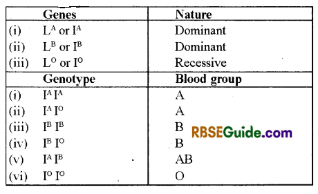
ABO System
General
It was discovered by Landstenier (1900).
According to it, there are two types of natural antigens on the RBCs namely A and B.
Similarity, two types of natural antibodies in the plasma namely anti A or a and anti-B or b
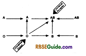
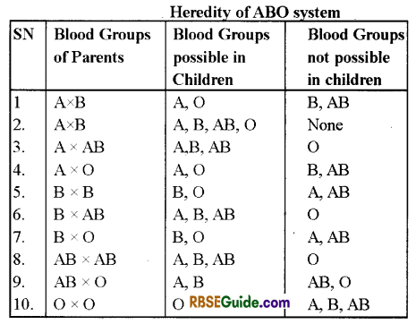
According to .ABO system, human blood is of four types as per table
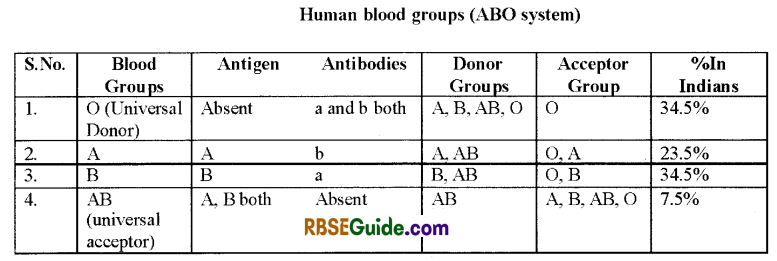
Group ‘O’ blood can be donated to persons with any other blood group and hence ‘O’ group individuals are called ‘Universal donors’. Persons with ‘AB’ group can accept blood from persons with AB as well as the other groups of blood. Therefore, such persons are called ‘Universal recipients’.
Genetics of ABO System
Bum Stein (1924-25) First explained that ABO blood groups in human beings are hereditary character.ABO system is controlled by three genes. Hence, it is an example of multiple gene inheritance.
![]()
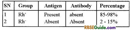
Rh System
General
Landsteiner and Weiner (1940) discovered Rh factor in Rhesus monkey. Rh antigen is found on the surface of RBC. Such individuals are called Rh positive (Rh+) and those without this antigen are called Rh negative (Rh–).
Rh antigen is, also called as Rh factor. It is controlled by dominant gene.
If Rh+ blood is transfused to the Rh person than it produces agglutinin. It is called Isoimmunization.
According to it, human blood is of two types-
Erythroblastosis foetalis
This disease occurs when husband is Rh+ and wife is Rh–. In this case, the foetus will be Rh+.
Rh antigens of the foetus do not get exposed to the Rh-ve blood of the mother in the first pregnancy as the two bloods are well separated by the placenta. However, during the delivery of the first child, there is a possibility of exposure of the maternal blood in small amounts to the Rh+ blood from the foetus. In such cases, the mother starts preparing antibodies against Rh antigen in her blood.
In case of her subsequent pregnancies, the Rh antibodies from the mother (Rh–) can pass into the blood of the foetus (Rh+) and destroy the foetal RBCs. This could be fatal to the foetus or could cause severe anaemia and jaundice to the baby. This condition is called Erythroblastosis foetalis
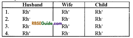
This can be avoided by administering anti-Rh antibodies to the mother immediately after the delivery of the first child. Baby suffering from this disease is called “Rhesus baby. Generally they born before stipulated gestation period and have blood deficiency complete blood transmission of new born can be done to save life of child. New born can also be saved by giving a mother Rh- antibody injection just before 72 hours of delievery.
Blood clotting or Blood coagulation
General
Blood exhibits coagulation or clotting as result of an injury or trauma. This is a protective mechanism which prevents excess loss of blood. Factors which induce clotting are known as Inducer and which inhile mogationis function clotting is called anticoagulant. In clotting, fibrinogen thrombin, platelets and total 13 factors are involved. This process occur in three steps-
1. Formation of prothrombin- Normally these factors remain inactive and become active on bleeding. The platelets break on air contact and release inactive thromboplastin. It forms active thrombin in presence of Ca++, factors VII and Factors VII and X and Ca++together called prothmbin activator.
2. Conversion of Prothrombin into Thrombin- Prothrombin is a protein found in plasma Thrombin activator converts it into Thrombin.
3. Conversion of Fibrinogen into fibrin and formation of Clot Fibrinogen is a soluble protein found in plasma and it changes into fibrin with the help of thrombin which is a non soluble and fibrous protein. These fibrin monomers soon assemble and form a net at the wound. This network with entangeled blood cells form a clot.
The liquid oozes out of the clot which is called as serum. The semm is plasma without fibringen. Normally, clotting takes about 6-10 minutes.There is no blood clothing inside the blood vessels because the blood contains an anti-coagulant called heparin
![]()
Cascade theory
Blood clothing can be explained with the help of cascade theory. The 13 clotting factors participate as follows-
- Intrinsic factors
- extrinsic factors
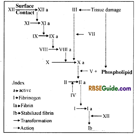
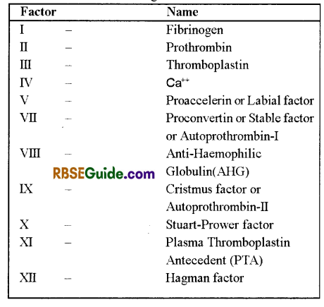
The names of 13 Clothing factors are as follows-
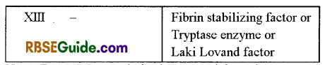
Note: Factor VI (Accelerin) is removed from the sequence as it is an anctive form of factor V.
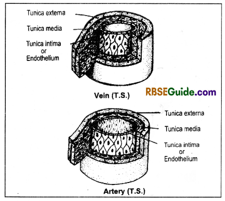
Blood vessels
General
These are the part of circulatory system. They transports blood. There are three kinds of blood vessels in system
- Arteries
- Veins
- Carallaries.
- Thin arteries are called arterioles and veins as vanules.
- The various arteries form arterial system & the various veins form venous system.
- The walls of the blood vessels are made up of 3 layers
- Tunica externa or adventitia
- Tunica media
- Tunica interna or intima or endothelium.
- The walls of arterioles & veinules are made up of only endothelium.
- The blood capillaries which supply blood to the walls of blood vessels are called as vasa vasorium.
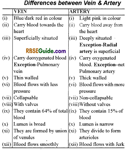
Differences between Vein & Artery
Heart
External structure of Heart.
The heart is situated in the thoracic cavity in between the lungs obliquely towards left side.
The anterior end of the heart is broad & it is called as base. The posterior end is narrow & it is called as apex.
The heart is covered by pericardium.
There is a pericardial cavity between the pericardium & heart which is occupied by pericardial fluid. This fluid protects the heart from shocks & keeps the heart moist.
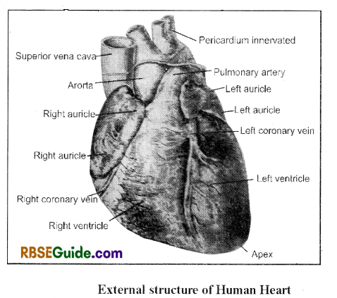
The walls of the heart is consist of three layers-
- Epicardium – outermost
- Mesocardium – middle layer & it is made up of Cardiac muscles.
- Endocardium – Inner layer
![]()
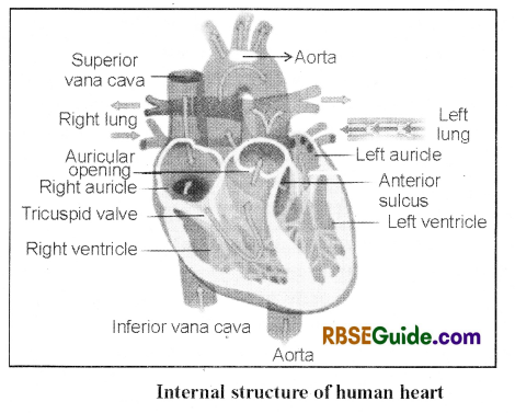
The anterior part of the heart is called as auricular part & the posterior part is called as ventricular part. There is a coronary sulcus between these 2 parts.
The auricular part is smaller & less muscular. It is divisible into 2 equal left & right auricles with the help of an interauricular septum.
During embryonic stage, the interauricular septum bears an oval pore, the foraman or alis. This pore get closed later & its scar is called fossa ovalis.
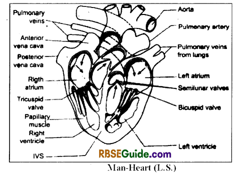
The interventricular septum is visible from outside ventrally. A coronary artery is situated along the interventricular septum.
The left auricle receives two pulmonary veins through a single slit-like & valve-less aperture. They bring oxygenated blood. In human beings, both the pulmonary r eins open through 4 small apertures into the left auricle.
The left auricle opens into a left ventricle through an aurieulo-ventricular aperture which is guarded by a bicuspid or mittral valve.
The left ventricle opens into left systemic arch and the opening is guarded by three semi-lunar valves.
The right auricle receives two precavals & one postcaval which open through separate & valve-less apertures.
The ventricular part is larger & more muscular. A oblique inter ventricular septum dir ides the ventricular part into unequal left & right ventricles.
The right auricle has a membranous eustachian valve and a sinu-auricular node (S-Anode). In addition, it receives a coronary sinus. It’s opeing is guarded by a Thebesian valve.
The right auricles opens into right ventricle and this opening is guarded by a tricuspid valve. The right ventricle leads into a pulmonary arch & it’s opening is guarded by 3 semi-lunar valves.
Both the pulmonary arch & systemic arch cross each other & remain connected with the help of ligamentum arteriosum. During embryonic stage, both the pulmonary & systemic arches remain connected by a ductus arteriosus which is also called as ductus Botalli. Later on this duct gets closed.
Both the ventricles are provided with papillary muscles which form pocket-like columnae cornae. Thread-like chordae tendinae remain attached betw een the papillary muscles & the bicuspid tricuspid valves. The chordae tendinae & the papillary muscles prevent reverse opening
of the valves.
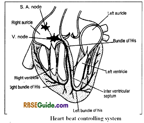
There is an aurieulo-ventricular node (A-V node) in the tip of the interventricular septum. The AV node gives out two bundles of His which further divide to form Purkinje’s. fibres.
![]()
Working of Heart
The heart functions like a pump & it keeps the blood circulating in the whole body.
The rhytlieric contraction of the heart is called as heart beat. Each heart beat has two parts-
- Systole
- diastole
During diastole the heart receives blood from the veins & during systole it pumps the blood into the arteries.
The heart contracts alternately i.e. when the auricular part contracts, the ventricular part relaxes & vice versa.
The number of heart beats in adult man is 72 per minute.
Mechanism of heart beats
The heart beats begins by the self contraction of the S-A node. Hence, S.A. node is also called as pace maker.
The S. A. node consists of cardiac muscles and nerve cells. Hence, the heart beat begins by the contraction of the cardiac muscles Therefore, the heart is also called as myogenic.
Firstly, the S. A. node contracts, as a result both the auricles contract. Soon the stimulus of contraction get transmitted to the A. V. node & from the A. V. node to all over the ventricles with the help of bund le of His & Purkinje’s fibres. It results in ventricular contraction.
Cardiac cycle
Each heart beat includes a systole and a diastole. The systole consisits of both the arterial and ventricular contractions. The human heart beats 72 times per minute, out of which the atrial systole lasts about 0.1 second and the diastole takes about 0.76 second (duration of each heart beat is 0.86 second).
The sometimes after the beginning of the atrial diastole, ventricular systole begins and it takes about 0.38 second. Since the aterial diastole takes a longer time, whole of the V. systole and most of the V.
diastole coincides with aterial diastole. Remaining part of the V. diastole coincides partly with auricular systole and partly with the early auricular diastole. The duration of ventricular diastole is 0.48 second.
![]()
The cardiac cycle is divided into two parts Ventricular sy stole and Ventricular diastole.
(1) Ventricular systole-During this phase, the blood from the ventricles enters into the arteries. It is further om divisible into 3 phases
(i) Isometric phase – This is the first part of the V. systole in which ventricular pressure increases. It results in the closing of A-V valves.
(ii) Period of Maximum Ejection – The semi lunar valves are forced open and the blood begins to pour out of the ventricles. The contraction of the ventricles continues and there is graduall decrease in V. blood volume.
(iii) Period of Reduced Ejection- During last part of the V. systole, the rate of blood ejection decreases and the V. pressure is minimum. The arterial pressure also falls during this period.
(2) Ventricular diastole : It includes 5 phases viz.-
(i) Protodiastasis-It is the transiet period between the systole and the diastole.
(ii) Isometric relaxation – At the end of systole. V. relaxation begins suddenly. There is closing of semilunar valves. During this period ventricular pressure falls.
(iii) Period of Rapid filling-The high pressure in the auricles pushes open the auriculoventricular valves and allow’ blood to flow rapidly into the ventricles.
(iv) Period of Slow filling-During this phase a small amount of blood normally flows into the ventricles.
(v) Period of last Rapid filling – It is the last part of the V. diastole during which atria contract and give an additional trust to the inflow of blood into the ventricles.
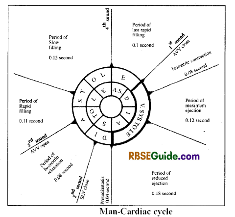
Heart Sounds & Pulse
The heart produces 4 sounds while functioning which are called as heart sounds viz.- I, II, ID & IV. Out of them 2 sounds are main & are called as classical sounds-
I, II…… ,I, II ………
The heart sounds can be beared with the help of a stethoscope.
First sound is dull & prolonged like L-U-B-B. It is caused due to closing of bicuspid & tricuspid valves. It concides with the beginmg of systole.
Second sound is short & sharp like DI F, It is caused due to closing of semi-lunar valves. It concides with the begining of diastole.
Third sound is a slow sound. It is caused due to opening. of bicuspid & tricuspid valves and sudden rush of blood
into the ventricles.
Fourth sound is very slow’ sound which is caused due to contraction of the auricles. It occurs just before the 1st sound.
![]()
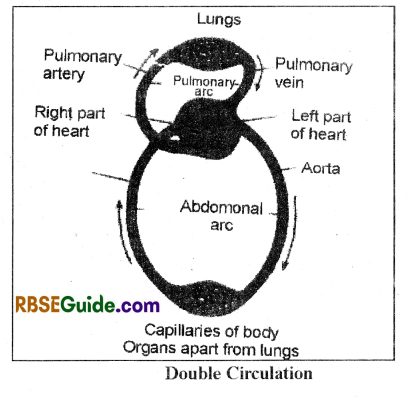
Double Blood Circulation
Physiologically, the heart is divisible into a right part & a left part. The right part contains deoxygenated blood and the left part contains oxygenated blood. In this type of heart, the blood flow’s twice through the heart before body circulation which is called as double circulation.
Significance It keeps the oxygenated and deoxygenated blood separate i.e. prevents mixing of oxygenated & deoxygenated blood.
It is made by Lymph, Lymph vessel and Lymph nodes.
Lymph
Lymph flows in fluid form in Lymphatic system. Blood gets filtered in blood capillaries due to blood pressure, that is called as Lymph.
It is colourless translucent & alkaline vascular tissue.
It is similar to plasma but the lymph is without RBC & platelets.
The lymph has less amount of protein, Ca++ & Phosphorus than the plasma.
The lymph is a carrier of nutrients, hormones etc.
It contains specialized lymphocytes which are responsible for immune responses of the body.
Lymph vessels
The lymph vessels are similar to the veins which carry lymph.
In lymph vessels, the lymph always flow from the organs towards the heart. They have less pressure than veins.
Lymph vessels are lined by endothelial cells and have a thin layer of smooth muscle, and adventitia that bind the lymph vessels to the surrounding tissue.
Main lymph vessels are as follows-
(i) Thoracic Duct
The main vessel of the lymphatic system, passing upwards in front of the spine and draining into the left innominate vein near the base of the neck. The thoracic duct is the largest lymphatic vessel of the lymphatic system of the body. It is approximately 40 cm in length in adults, and approximately 5 mm in width at its abdominal origin.
(ii) Right Lymphatic duct
The right lymphatic duct drains lymph from the right upper limb, right side of thorax and right halves of head and neck. The thoracic duct drains lymph into the circulatory system at the left brachiocephalic vein between the left subclavian and left internal jugular veins.
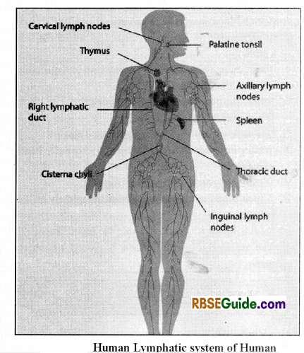
Lymph Nodes
There are lymph nodes at many regions of the lymph vessels. These nodes have network of lymphocyte granules.
They are situated close to the large blood vessels in head, neck, arm pits, groins etc.
Tonsils are also lymph nodes.
Functions of Lymphatic system
(i ) Lymph acts as intermediate between blood & the tissues. It takes digested food & O, from the blood & provides to the tissues. Similarity, it takes excretory substances, hormones etc from the tissues & convey them to the blood.
(ii) Lymphatic system helps in the circulations of absorbed substances particularly fatty acids & glycerol.
(iii) The digested fats are absorbed into the lacteals of the ileum.
(iv) The WBC found in the lymph destroy the Germs.
![]()
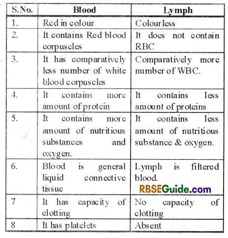
Differences between blood and lymph
Diseases related to heart and circulatory system
Hypertension or High Blood Pressure
The BP in human beings is expressed as Sp DP mm of Hg. The normal value is 120/80 mm of Hg.
An increase in SP/DP Both is called as hypertension.
It adversely affects brain, kidneys & heart.
Coronary Artery Disease
- It is also called as Atherosclerosis. It is caused by deposition of calcium, fats, fibrous material in the arteries supplying blood to the heart muscles.
- As a result, the artery lumen becomes narrow.
Heart Failure
- It is also termed as congestive heart failure. The heart failure is different than heart attack, his heart attack, the heart beat stops suddenly.
- The heart failure is usually caused by insufficient blood supply to the heart muscles.
- The main Symptom is congestion of the lungs.
Angina
- It is normally caused due to coronary thrombosis. As a result, the Cardiac muscles fail to get proper oxygen and nutrients.
- There is severe pain in the chest migrating towards left arm.
- Also termed as angina pectoris.
Myocardial Infarction (MI)
- MI is also called as heart attack.
- It occurs when the coronary artery gets blocked, normally because of coronary thrombosis.
- The cardiac muscles get damaged because of less blood supply and the heart fail to function with full capacity.
Valvular Disease
It is because of malfunctioning of the cardiac valves, as a result the blood starts flowing in opposite direction.
There is mixing of oxygenated & deoxygenated blood.
Ventricular Fibrillation:
In this disease, the vantricular muscles contraction disorganized. The quivers instead of pumping.
Paricardiatis:
It is caused due to bacterial infection in the pericardum. There is inflammation in the pericardium and increased amount of pericardial fluid. It causes more pressure on the heart which may be fatal.
Coronary Thrombosis:
The intravascular blood clot is called as thrombus & its formation is called as thrombosis. When the thrombus forms in the coronary artery, it is termed as coronary thrombosis. It may result in heart attack.
![]()
Heart Block:
It is caused due to malfunction of Bundles of His. As a result, the impulses generate in the S-Anode fail to spread all over the ventricles. Hence, there is no/poor ventricular contraction.
Rheumatic Heart Disease:
It is caused by the infection of Streptococcus viridines bacteria. The heart valves do not work properly and cardiac muscles become weak.