Rajasthan Board RBSE Class 12 Biology Notes Chapter 26 Man-Nervous System
General
Various vital activities of the body are controlled & coordinated by two systems:
- Nervous System
- Endocrine System
This chapter deals with the study of the Nervous System.
The nervous system is divided into 3 heads:
- Central nervous system – It includes the brain & spinal cord.
- Peripheral nervous system – It includes cranial & spinal nerves.
- Autonomous nervous system – It includes the sympathetic & parasympathetic nervous system.
![]()
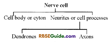
A nerve cell or Newron is the structural and functional unit of Nervous System.
The space in between the neurons is occupied by neuroglial cells in the nervous system.
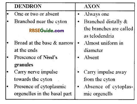
The cyton has a large & round nucleus, minute neurofibrils, cytoplasmic organelles & many Nissl’s granules. The Nissl’s granules are ribosomes & made up of RNA & proteins
However, the centrosome is absent. Hence, the nerve cell never divides.
Nissl’s granules are the groups of lamillae which are attached with ribosomes. They synthesize proteins actively.
The nerve cells increase in size along with body’s growth.
The neurons are of 3 types on the basis of cell processes viz.
(i) Unipolar:
It has only one axon.
They are found in invertebrates & vertebrate embryos.
(ii) Bipolar:
It has one axon & one dendrone
They are found in eyes, cochlea & olfactory epithelium
(iii) Multipolar:
It has one axon & many dendrites
Most vertebrates have multipolar neurons On the basis of the structure of axon, the neurons are of two types
(a) Myelinated or Medullated:
The axon is bounded by a myelin sheath
(b) Non-myelinated or Non-medullated:
The axon is not bounded by myelin sheath
Functions of Nervous System:
- It receives and coordinates activities of various organs of the body.
- It informs the body of man about the changes in the external environment through the receptors.
- It conducts or transmits messages to various parts of the body.
- It also regulates many involuntary activities such as breathing, heartbeat, movement of food in the alimentary canal etc.
- It controls all voluntary’ activities of our body such as running, speaking etc.
- The nervous system works like a complete unit in the body by coordinating different activities, it helps to maintain steady state of the body.
![]()
Brain
General
The brain is the softest body organ which remains protected by Cranium (Skull)
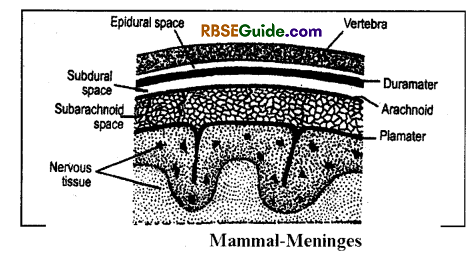
In addition. The brain & spinal cord are covered by three protective membranes which are called as meninges.
There is one menin in fishes (Piamater or menix Primitiva); 2 meninges in amphibians (Piamater & dura mater) and three meninges (Piamater, arachnoid & duramater) in mammals
Dura mater is outermost, thick & tough, and made up of collagen fibres.
There is an epidural space between vertebral column & dura mater
Arachnoid is the middle menin which is highly vascular
There is a sub dural space between arachnoid & duramater which is full of CSF.
The piamater is thin . elastic, vascular, & inner most layer which provides nourishement.
There is a sub arachnoid space between arachnoid & piamater which is full of alkaline CSF.
At many places, the piamater & arachnoid fuse to form leptomenin
Viral/bacterial infection in the meninges causes meningitis.
Cerebro – Spinal Fluid (CSF)
A lymph-like fluid is secreted and comes out as filtrate from the choroid plexus. It is called cerebrospinal fluid (CSF).
The CSF contains nutrients such as amino acids, glucose, chlorides, bicarbonates, Na+, K+, Ca++, P04– S04–, etc.
A healthy person has 150 ml of CSF.
The CSF flows from anterior to posterior, travels through subarachnoid cavity and finally enters into the blood through the blood capallaries of arachnoid.
Functions of CSF:
- It protects the brain & spinal cord from injuries and also keeps the brain moist.
- It provides nutrients to the brain & spinal cord.
- It helps in the exchange of 02 & C02 between blood & brain.
- It is used to diagnose brain related diseases.
- Increased amount of CSF also caused menigititis.
![]()
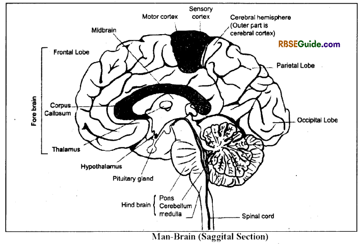
Structure of Brain
Human brain is divisible into 3 parts
- Forebrain
- Cerebellum
- Brain stem
Forebrain
In human beings, the forebrain includes two parts-
(A) Cerebral hemispheres or cerebrum
(B) Diencephalon
(a) Cerebrum
It forms 2/3 part of the brain and a longitudinal fissure divides it into two hemispheres.Both the hemispheres remain connected internally by a corpus callosum.
Each cerebral hemisphere consists of five lobes-
- Frontal lobe
- Parietal lobe
- Temporal lobe
- Occipital lobe
- Insula lobe
The insula lobe is not visible externally.
The frontal lobe is the largest. A central sulcus separates the frontal and pariental lobes which is also called as the fissure of Ronaldo. Similarly, a lateral cerebral fissure separates the frontal and the temporal labes which is also called as the fissure of Sylvius. The parietal and the occipital lobes remain separated by a parieto-occipital fissure.
The outer 2 to 4 mm thick part of the cerebrum is called as cortex or grey matter. It consists of 10% of the total neurons of the brain. The cortex exhibits distinct gvri (ridges) and sulci (furrows). These gyri and sulci increase the surface area of the cortex by 3-times. The inner mass of the cerebrum is called as white matter.
Functions of the cerebrum
(i) Frontal lobes: They control memory, reasoning, thinking, determination, emotions, judgment, planning, experience, concentration, will power and personality. They also control voluntary’ muscles and interpret many sensations.
(ii) Parietal lobes: They interpret sensory perception of touch, pain, cold, heat etc. They also control speech and handicraft. They also associated with the understanding of thoughts.
(iii) Temporal lobes: They interpret and decode sound. In addition to language comprehension, they also interpret smell and auditory sensations.
(iv) Occipital lobes: The interpret and decode visual sensations particularly sensations concern with colours and shapes. They also control eye movements.
(v) Insula lobes: The coordinate memory and other parts of the cerebrum.
(b) Diencephalon
It is divisible distinctly into epithalamus, thalamus and hypothalamus.
Thalamus forms 80% of the diencephalon and it functions as relay centre of the sensory’ perception of taste, touch, hearing, vision, pain, heat etc. It interprets these sensations and sends motor drives concerning to the voluntary movement.
![]()
The hypothalamus includes about 12 large nuclei and it is divisible into four parts:
(i) Supraoptic part : It is situated just above the optic chiasma. It’s axons enter into the pars nervosa of the pituitary gland.
(ii) Tuberal part: It is the broadest and the middle part. It is attached with the pituitary through a stalk called the infundibulum. It has a projection, the tuber cineriuni around the infundibulum.
(iii) Mammillary part : It is the posteriormost part which bears two small and round projections, the mamillary bodies. It controls reflex actions concern to smell.
(iv) Preoptic part: It is a small part which is situated anterior to the supraoptic part. It control some autonomic reflex actions.
Function of the hypothalamus:
- It controls coordination between psyche & body and neurons & endocrine systems.
- It has centres of hunger, thirst, satiety, and temperature control.
- It includes nuclei secreting oxytocin, and vasopressin hormones.
- It controls diumal rhythm and sleep
Mid Brain
- It is the middle part of the brain and also called as mesencephalon.
- It consists of two parts-
(a) Corpora quadrigemina
- It includes four optic lobes which are collectively called as Corpora Qudrigemina.
- Out of 4 lobes, two lobes are surerior optic lobes and two are inferior optic lobes.
- The optic lobes remain joined to the dorsal surface of midbrain with the help of Tactum.
- The optic lobes receive stimuli related to vision & hearing.
(b) Cerebral peduncle
- It is also called as Crura Cerebri.
- It is a bundle of nerve fibres that connects cereberal cortex to the spinal cord & other parts of the brain.
Hind Brain
It is posterior or terminal part of the brain. It consists of three parts, cerebellum, pons varolli and medulla oblongata.
(a) Cerebellum: It is the second largest. It part consists of two lobes. Dorsal side of cerebellum is well developed. It’s two main functions are-
- Maintenance of posture or equilibrium.
- Coordination of muscular activities.
(b) Pons Varolli: It lies just above medulla oblongata and below cerebral peduncle. It joins both lobes of cerebellum and also other parts of the brain. It is made up of white matter.
It contains Pneumotaxis centre for regulation of breathing. It also controls chewing, saliva secretion, release of tears and eye movements.
(c) Medulla Oblongata: It is the posterior-most part of brain. It is triangular and situated between pons and spinal cord. Its cavity is called nietacoel and medulla continues as spinal cord. Functions of medulla oblongata are :
- It has controlling centres of bleating, coughing, swallowing etc.
- I has controlling centres for heart beats, blood pressure, peristalsis in alimentary canal & other involuntary activities.
![]()
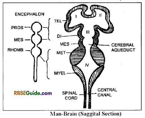
Brain Ventricles
The mammalian brain is hollow and the cavities are called as ventricles. These ventricles are lined by ependymal epithelium and are lull of cerebrospinal fluid (C.S.F.).
The brain has 4 ventricles viz.-I, II, III & IV.
I & II ventricles are found in the cerebral hemisphres and are also called as paracoels
Each paracoel forms a rhinocoel in the olfactory lobe which is also called as zero ventricle.
Both the paracoels open into a third ventricle through a foranian of Monro.
III ventricle is also called as diocoel & it is found in the roof of the diencephalon.
The 3rd ventricle opens into IV ventricle through a narrow duct, the iter or aqueduct of Sylvius. The iter is found in the cerebellum.
IV ventricle is also called as nietacoel & it is found in the medulla.
There are two choroid pluxes in the ventricles viz.-
- Anterior choroid plux: It is found in the roof of the diocoel .
- Posterior choroid plux: It is found in the roof of the metacoel. The choroid plux consists of piamater & network of blood capillaries.
The anterior choroid plux secretes CSF and the posterior choroid plux absorbs CSF & secrete it into the subarachnoid space. There are three apertures in the posterior choroid plux to absorb the CSF viz.
- Foraman of Magendie: One
- Foramen of Luschka: two
There are arachnoid villi in the subarachnoid space. These villi absorb CSF & return it into the venous blood.
![]()
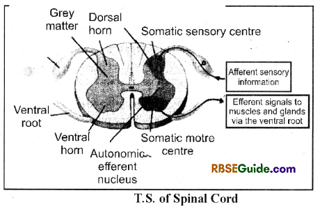
Spinal Cord
Structure
It originates from the medulla & it comes out of the skull through a foraman of Magnum
It is situated in the neural canal of the vertebral column.
In the tail, it terminates & it’s last part is called as filum terminale.
It is covered by 3 meninges but the Ilium terminale is covered by piamater only.
It has a dorsal fissure and a ventral fissure.
It’s central cavity is called as neurocoel which originates from the metacoel.& it is full of CSF.
Structurally, it has outer white matter & inner grey matter.
The grey matter is butterfly or H-shaped & it forms 2 dorsal horns & 2 ventral horns at the point of origin of the spinal nerves.
One dorsal horn & a ventral horn of one side unite to form a spinal nerve.
The dorsal horn acts as a sensory – root & ventral horn as a motor root.
The grey matter divides white matter into 4 parts, out of them one is dorsal funiculus, one is ventral & two are lateral funiculi.
Functions of Spinal Cord :
- To cany reflexes below neck.
- To carry sensoiv message (stimulus) from skin and muscles to Brain.
- Response from brain to hand, legs, & body.
- It acts as centre for spinal reflex.
- It helps brain in its all functions.
- Cerebrospinal fluid present in it, protects spinal cord from external shock or injury.
- Permeability or movement of different substances and maintaining appropriate pressure in brain.
- Control and co-ordinate various involuntary actions.
Peripheral Nervous System
The nerves originating from brain and spinal cord are included in peripheral nervous system, i.e. it includes Cranial nerves arising from brain and Spinal nerves arising from the spinal cord.
![]()
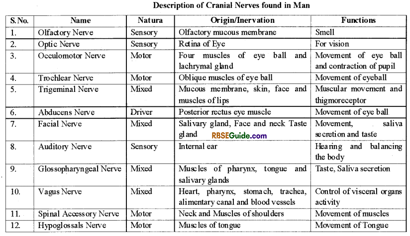
Cranial Nerves
There are 12 pairs of cranial nerves in man. These nerves arise from ventral side of the brain. They may be sensory, motor or mixed, it depends on their function
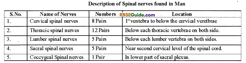
Spinal Nerves
- These nerves arise from spinal cord.
- There are 31 pairs of spinal nerves.
- All spinal nerves are of mixed type as they contain both sensory and motor neurons.
- Every spinal nerve pass out through each-two vertebrae of vertebral column through inter-vertebral foramina.
- On the basis of their origin, the spinal nerves are named as follows
Autonomous Nervous System
It is discovered by Langley. It is completely motor and without control. It provides nerves to involuntary muscles and glands.
It is formed by ganglions and chains of a pair of nerves, which is located on both lateral side of spinal cord.
It is mainly a stimulatory system, which control the involuntary activities of visceral organs.
It is of two types :
- Sympathetic nervous system,
- Parasympathetic nervous system
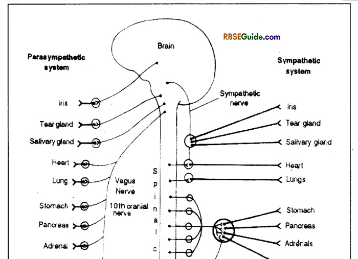
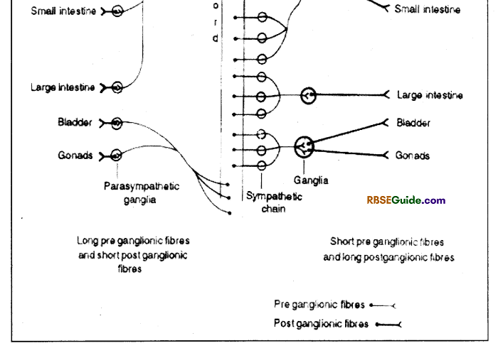
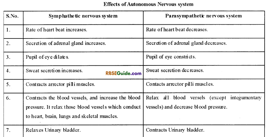
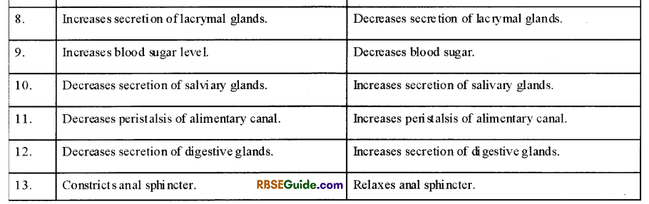
Sympathetic nervous system prepare the body for conditions arises in emergency and parasympathetic nervous system establish the body in normal position at the end of emergency. Both the systems function antagonistically. The opposite effects of both these system’is given in table .
The emotions have too much impact on autonomous nervous system such as -unhappiness, fear, sexuaL stimulation etc.
![]()
Reflex Action
General
A reflex action is a rapid automatic response to a stimulus without conscious thought in the body. The response is instantaneous and involuntary’.
There is no control of brain on reflex actions.
For example-
(i) A person withdraws hand instantly on touch to a pointed pin or hot utensil.
(ii) Salivation in the mouth on right or smell or thought of good favourite food.
Reflex actions are to two types-
- Simple or unconditional reflex action
- Acquired or conditional reflex action
Unconditioned Reflex action
They are natural or congenital.
Learning is not required.
Examples-
- Instantly closing of eye lids when any object comes close.
- Instantly coughing when any food particles any liquid enters into trachea.
- Contraction of pupil in strong light.
Conditioned or Acquired Reflex action
This reflex action needs leaming/training or experience.
Russian Scientist of physiology’ Evan Pavlov explained it by his experiment on dog.
Experiment on Dog-Pavlov start ringing a bell just before the food is given to Dog. Lateron, he observed that there is salivation in Dog’s mouth on ringing bell even without giving food.
Examples-
- We instantly brake our vehicle when any object comes in front of it all of sudden.
- Tying of shoe laces even though wv are talking to somebody else.
- Running away of dog, when we just bend down towards ground as dog pretends that we are picking stone to throw on him.
- Standing up of student as soon as teacher enters into class.
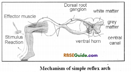
Mechanism of Reflex Action
General
Fibres of spinal cord and motor fibre of ventral root play special role in reflex action.
For example – pin prick on the skin stimulate sensations. Which stimulate dendrites of somatic sensory’ fibres. These fibres transmit stimulus to the neurons in the dorsal ganglia of spinal nerve.
Terminal node of these cells send stimulus to dendrites of nearest motor nerves. Here sensory induction becomes motor stimulus. Axons of motor cells are fibres of ventral root and they carry stimulus up to legs, where contraction in muscles and produce movement of legs.
These actions are very fast and in total it is called reflex action.
Spinal Reflex Arch
The nerve path way involved in a reflex action is called a reflex arch. It includes following 5 parts:
- Sensory organ: Receptors on the body which receive external or internal stimulus.
- Sensory Nerve: Sensory nerve carries impulse from sensory organs to spinal cord.
- Nerv e centre of spinal cord or Interneurons Centre of actions.
- Motor nerve: Transmit, impulse away toward spinal cord.
- Effector organs: These organs complete their actions with response to impulses received through motor nerve.