Rajasthan Board RBSE Class 12 Biology Notes Chapter 28 Man-Reproductive System
General
All animals exhibit reproduction as a fundamental property of living being to increase the number of it’s own kind. The higher animals perform only sexual reproduction which is with the help of special reproductive organs.
Human beings are unisexual or dioceous animals i. e. male & female reproductive organs are found in separate individuals.
The human beings exhibit sexual diamorphism i.e. male & female can be distinguished on the basis of external characters.
In sexual reproduction, the animals produce male & female gametes. They fuse to form zygote which is called as syngamy. The zygote finally develops to form a completely new animal.
The organs forming the gametes are called as primary organs and the rest organs are called as secondary or accessor) organs.
Females have more responsibility than males in reproductive process because many functions related with preproduction carried in females such as ovum formation reception of sperm during copulation, provide proper arrangement for pregnancy, provided nutrition to embryo etc.
Secondary sexual characters in man are presence of moustache and beard and heavy voice while in females (woman) feminine voice, soft skin, enlarged breasts, pelvic broadening with fat deposition are main characteristics.
![]()
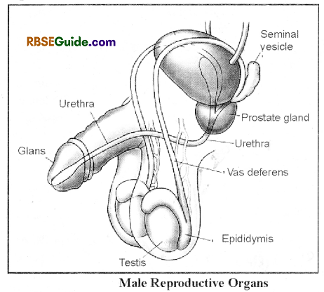
Male Reproduction System
General
The male reproductive system consists of primary reproductive and accessory reproductive organs. Primary sex organs are a pail – of testes or male gonads. Accessory reproductive organs are mainly scrotal sac, epididymis, vas deferens, penis, prostate gland and cowper’s glands.
Testes
Adult man has a pair of testes (oval bodies suspended outs the body in scrotum). The wall of tests is an clastic covering of thin and hair)’ and thick inner subcutaneous layer of unstriated muscle fibres which is known as Dartos muscle.
In each half of scrotal sac, a tuft of rod shaped striated muscles fibres is found. It conneccts subcutaneous layer with abdominal subcutaneous muscle and known as Cremaster muscle. Each cavity of scrotal sac is connected to the abdomial cavity with the help of a white Inguinal canal.
In adult mammals, testes are ex-abdominal which arc found outside the abdominal cavity in the testes sacs or scrotal sacs. The spermatogenesis in the testes-needs less temperature and the temperature of the scrotal sacs is 33 to 35°C which is 2 ° to 4° C less than the body temperature.
The testis descends into the scrotal sac through an inguinal canal. The testicular artery, testicular vein & the testicular nerve also descend into the scrotal sac in the form of a spermatic cord.
Each testis is attached in the scrotal sac with the help of a gubernaculum.
Both the spermatic cord & the gubernaculum are pro-vided with elastin fibres.
If the testes fail to descend into the scrotal sacs, the spermatogenesis gets arrested due to high temperature which called as cryptorchidism.
![]()
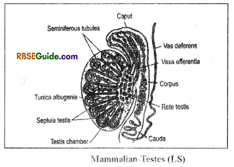
Internal Structure (Histology) of Testis
Testis is covered by tunica albugenia.
Internally, the testis is divided into an outer large chamber & an inner small chamber with the help of a septum called mediastium testis.
The inner small chamber of the testis has a network of narrow tubules which is called as rcte testis.
The seminiferous tubules open into the rete testis and the rete testis in turn open into vasa efferentia which are 15 in number. These vasa efferentia open into the caput part of the epididymis.
The larger outer chamber of the testis has transverse septa, the septula testis which divide it into 200 small chambers. Each small chamber has 2 to 3 seminiferous tubules.
The seminiferous tubule is a structural & functional unit of testes.
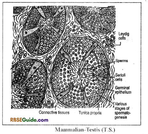
The space in between the seminiferous tubules is occupied by the interstitial tissues which contains special interstitial or Leydig cells. These cells secrete androgen hormone.
Each seminiferous tubule is bounded by tunica propria & it is lined by germinal epithelium.
The seminiferous tubule contains various stages of spermatogenesis in addition to the Sertoli cells
The sertoli cells are also called as supporting cells or nutritive cells or nurse cells or mother cells or subtentacular cells.
The Sertoli cells are found in all mammals & they are columnar. They provide support & nutrition to the sperms. The developing sperms remain embedded into these cells. The Sertoli cells also secrete inhibin hormone which inhibits FSEI.
The sertali cells also secrete Anti-Mulerian hormone which keeps the Mulerian ducts inactive.
![]()
Epididymis
It is thin, highly coiled and comma shaped tube about 6 meter long that leads into a vas deferens. Its coiled rings are adhered by connective tissues. It’s outer side is covered with thick muscular layer and inner side is lined with stratified epithelium.
It is attached to the inner surface of the testis & has 3 parts:
(a) Caput or globus major
(b) Corpus
(c) Cauda or globus minor
Globus major is a large cap-like structure which receives the vasa efferentia.
Globus minor is a small structure which opens into vas deferens.
The epididymis stores the sperms & it is the site for physiological maturation of the sperms.
Vas Deferens
It is about 45 cm. long & thin which receives the cauda epididymis tube. Inner surface of vas deferens is lined by pseudostratified columnar epithelium. Some of its cells secretes special fluid which keep inner path smooth for the movement of spermatozoa.
The vas deferens ascends into the body cavity through the inguinal canal.
The vas deferens forms a swollen part known as Ampulla at the posterior end of urinary bladder in the abdominal cavity. At this point a small duct of seminal vessel open in it.
Both the ducts unite of Vas deferens and seminal vesicle unite form an Ejaculatory duct which opens into Urethra.
Urethra
Ureter collect urine fromurinary bladder and on the way it units with ejaculatory duct, and form a urinogenital duct or Urethra. It is about 20 cm long and it passes through the penis and opens at the tip through urinogenital aperture.
Sex Accessory Glands
Three types of accessory glands are found in man. which secretes their secretions into the Urethra. These secretions are essential for viability and motility of spermatozoa. The secretions of accessory glands, epididymis, and sperms are collectively form semen. Following accessory reproductive glands are found in man
Prostate Glands
It is situated around the anterior end of urethra and opens into the urethra. Prostate gland secretes white fluid which form 25-30% part of semen. It is alkaline secretion and neutralize the acidic medium of the Vagina. This fluid contains Phosphatase, citrate, lysozyme, fibrinolysin, spermin etc.
It’s secretions activate the sperms and prevent the semon to coagulate.
In aged men the size of prostate may enlarged which create problem in urine discharge.
It consists of Four lobes-
- Ventral lobe-1
- Dorsal lobe-1
- Lateral lobes-2
![]()
Seminal Vesicles
- It is also called as uterus masculinus.
- There is a pair of seminal vasicles in man.
- The secretions of the seminal vesicle forms 70% part of the seminal fluid.
- The secretions of the seminal vesicle are alkaline, slimy & it’s pH is 7.4. It consists of fructose, ascorbic acid,
- prostaglandin & enzymes.
- The fructose provides energy to the sperms.
Cowper’s glands
- There is a pair of Cowper’s glands which are also called as Bulbo-urethral glands.
- They are small, round & yellow in colour and open into the middle part of the urethra.
- They secrete transparent &. alkaline fluid having pH 7.5 to 8. This fluid makes the urethra alkaline before the copulation
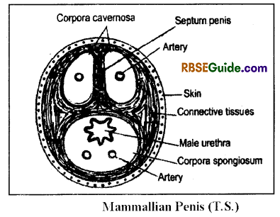
Penis
In man penis is suspended between the legs along with the scrotal sacs attached to the abdomen. It is cylindrical, erectile and highly vascularized eopulatory organ. It is covered by skin.
Body of penis is made up of three blood sinuses composed of filamentous, muscular connective tissues.T wo large are dorsolateral and one is ventrallv around Urethra.
Dorsolateral sinuses are called corpora cavernosa and ventral sinus as corpus spongiosum. Apex of penis is swollen and form glans. Gians penis is form of only corpus spongiosum. Covering of skin over glans is a cap like &. called as prepuce. It gets folded on the base of glans during copulation.
In normal condition blood sinuses remains empty and muscles are in contraction stage, at this time only urine can tlow outside through wrinogenital aperture.
mating, blood moves in the sinuses by penial artery, muscles relaxed and glans of penis becomes hard and swollen as maximum as possible. Glans become riacked from prepuce and it is now in the erection state. Penis is ready for deep penetration in the v agina of female. The semen ejaculated by penis into the vagina.
Semen
The fluid released by the male reproductive organs is called as semen. It includes sperms & a fluid.
It is a thick & milky fluid. It’s pi I is 7.45 and it’s density is 1.03.
The forceful release of semen from the male reproductive organs is called as ejaculation.
In human beings, 2 to 5 ml of semen is ejaculated at a time.
The number of sperms in the semen is 10 crore per ml.
It has seminal plasmin which acts as bacteriacidal.
The secretions of the prostate gland coagulate the spenn. In coagulated semen, the spenn movement is less.
Alter Sometimes, the fibrinolysin makes the semen liquid.
The prostaglandin found in the semen stimulates peristalsis in the uterus, as a result the sperms are sucked from the site of deposition into the uterus.
![]()
Female Reproductive Organs
One pair of ov aries are found in woman as a Primary reproductive organs. Accessory’ reproductive organs found in female are oviducts, uterus, vagina and vulva. Reproductive glands and mammary glands are also included as accessory organs.
Man – Reproductive System
Ovary
General:
- Ovaries are paired structures found in the abdominal cavity.
- Size of each ovary is 1.5 -3 cm long and 8 nun thick and shape is like an almond.
- It is found behind kidneys in the pelvic region.
- Ovary forms ovum and secretes female hormone, estrogen and progestrone.
Histology of Ovary
There is a pair of ovaries which are attached dorsallv in the abdominal cavity with the help of mesovarium. flic mesovarium originates from the visceral peritoneum.
The ovary has germinal epithelium inside the mesovarium which is made up of cuboidal germ cells.
The ovary has stroma made up of fibrous connective tissues. The peripheral part of the stroma is condensed which is called as cortex & the inner less dense part is called as medulla.
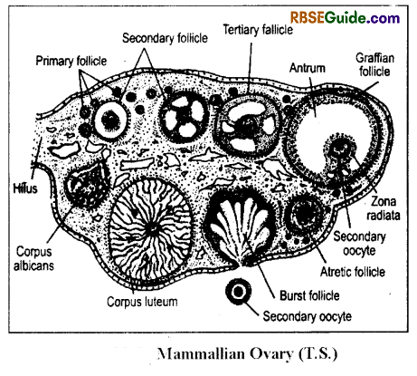
The cortical part of the ovary has ovarian follicles which are as follows
(i) Primary follicles:
They are formed during embryonic stage & begin to grow at puberty.
It has an oogonium surrounded by one layer of follicular cells.
(ii) Secondary follicles:
It has a primary oocyte surrounded bv 2 layers of follicular cells.
(iii) Tertiary follicles:
It has primary oocyte surrounded by 3 layers of the follicular cells.
(iv) Mature follicles or Graaffian follicles:
It is surrounded by a membrana granulosa which consists of two layers v.z.-theca externa & theca interna.
It has a fluid filled cavity, the antrum.
The mature follicle has a secondary oocyte which is attached to the membrana granulosa by a stalk. This stalk is called as germ hill or discus proligerous or cumulus oophoricus.
The Graffian follicle gradually migrate to the periphery & gets protruded.
The mature follicles bursts at the surface to release the secondary’ oocyte outside. It is called as ovulation.
(v) Atretic follicle:
Some times, the Graffian follicle fails to ovulate & degenerates gradually. The degenerating Graffian follicle is called as atretic follicle and the process is called as atretia.
It is due to deficiency of vitamin E & by hormonal imbalalnce.
(vi) Corpus luteum:
- The follicular cells of the burst follicles reorganize under the control of LH to form corpus luteum.
- It has yellow coloured luteal cells containing lutein yellow pigments.
- This structure performs endocrine function & secretes progesterone & inhibin hormones.
- There is a blood clot in the centre of the corpus luteum which is called as corpus haemorhagicum.
(vii) Corpus albicans
It there is no fertilization, the corpus luteum begins to degenerate. The degenerating coipus luteum is called as corpus albicans.
It is colourless & non-functional.
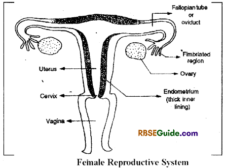
Oviduct
There is a pair of oviducts which originate from the Mullerian duct.
The anterior end of the oviduct modified to form ciliated fimbriated funnel or infundibulum. It’s mouth is funnelshaped which is called as ostium.The ovum enters into the oviduct through the oviducal funnel.
The middle part of the oviduct is called as Fallopian tube.
Its wall has unstriated muscles & ciliated internally. The Fallopian tube is the site for secondary maturation division, fertilization & embryonic development.
![]()
Uterus
The posterior part of the oviduct modifies to fonr The uterus is simplex in woman.
Uterine wall has three layers-
- E pirn atrium – Outer & made up of visceral peritoneum.
- Myomatrium – It is the middle layer which is made up of smooth muscles. It contains the longest smooth muscle.
- Endomatrium – lt is the innermost layer which has 2 layers viz.-stratum functional & stratum basale.
Vagina
The large, elastic & muscular tube that runs from cervix (terminal part of uterus) to outside is called Vagina.
The Vaginal opening is partialy closed by a thin membrane called Hymen. This hymen may be tom off due to physical labour, sexual contact and exercise etc.
Vagina provides path for menstrual discharge beside copulatory organ of female. It serves as a birth canal during the birth of the child. Two folds of tissue called vulva protect the vaginal and urethral openings.
Vaginal wall stores glycogen. The Lactobacilli bacteria present in the vagina makes mucous acidic by fermentation. This prevent vagina from infection.
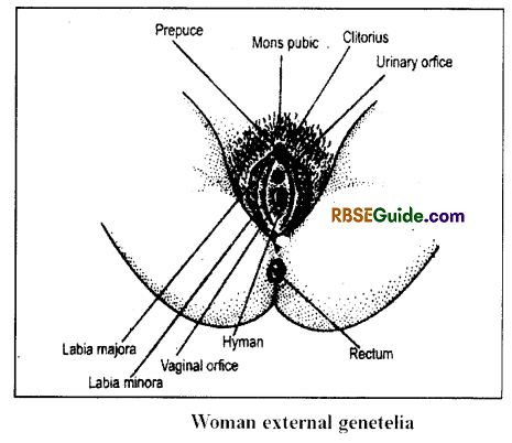
Vulva
External genitalia of woman is called vulva. It is situated just above the perinaeum is pelvic area.
Following structures are included in vulva :
(i) Mons pubis or Mo ns veneris : It is swollen fatty tissue covered by skin and situated above pubic symphysis
(ii) Labia Majora : A pair of transverse folds expanded from mons pubis to lower side and till back. It is tissue covered by skin and hair are found on its surface.
(iii) Labia Minora : There is a pair of small folds present inside the labia majora surrounding vestibule.
(iv) Clitoris : It is a sensory and erectile oigan situated at anterior comer of labia minora and below the mons pubis. This is homologus to glans of penis (intromittent organ of male).
(v) Vestibule : ‘Afissure like structure in the middle of labia minora is called vestibule. The urinary orifice and orifice of vagina are found. Below the clitoris is the urinary orifice or meatus is found as a small aperture. Below this is orifice of the vagina
is situated.
Sex Accessory Glands
There are two types of sex accessory glands-
Prostate Glands:
A pair of prostate glands open into the anterior part of the vagina. Their secretions make the vagina moist before copulation.
![]()
Bartholin Glands:
On both sides of the vaginal opening a pair of bean shaped glands are situated. These are Bartholin’s glands.
These glands secrete an alkaline and lubricating fluid, which keeps the vulva moist and facilitate sexual intercourse.
Breasts:
Breasts begin to develop at the age of puberty and finally become Mammary glands as accessory reproductive organs of female.
One pair of mammary glands found in women. These are found on the pectoral muscles on ventral side.
Each gland composed of connective tissues, 15-20 tubular partitioned lobules. Fatty tissue is found between these structures. Each lobule has proliferation of glandular tissues caused by the action of estrogen and progesterone.
Glands secrete milk as the nutrition of new born. Many small ductules unite and form lactiferous duct in each lobule. Such many lactiferous tubules open on the nipples independently.
Nipple is a knob like pigmented structure on each mammary gland. The area around nipple is darker (dark pigmented) and called as areola mammae. Each nipple has 15 to 25 pores for milk ejection.
Growth and function of mammary glands is controlled by somatotropin, prolactin, estrogen, progesterone and oxytocin hormones.
Milk contains fat, lactose, casein, lysozyme, calcium, vitamin and important immunoglobins.
Mother’s milk is a complete food for the neonatal and it develops immunity in the child.
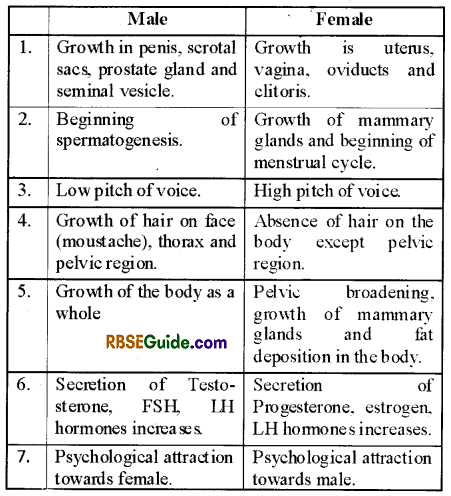
Onset of Puberty in Human beings
Maturation of reproductive organs in male and female human beings and development of reproductive capacity is called onset of Puberty.
In male it occurs at 14-16 years of age while in female it begins at the age of 12-14 years.
Growth of mammary glands and menarche (first time menstruation) is sign of puberty in human female.
Table Changes in Male and Female at Puberty