
Rajasthan Board RBSE Class 12 Biology Notes Chapter 25 Man-Excretory System
General
- Various metabolic activities in the body results in the formation of waste products, such as CO2 nitrogenous
- substances (ammonia, urea, uric acid) water etc.
- The removal of these metabolic waste products is called as excretion.
- The excretory’ system concerns with the removal of the nitrogenous waste products.
- Kidneys are the primary excretory organs.
![]()
Various Nitrogenous Waste Products
Nitrogenous waste products are formed by catabolism of proteins. They are as follows :
Ammonia
Catabolism of proteins release ammonia in all the organisms. It is highly toxic, hence should be eliminated soon.
The animals which excrete ammonia are called as ammonotelic.
Ammonia is very less soluble in water, hence needs more water in its excretion. Therefore only aquatic animals can afford to excretie ammonia.
Examples : Protozoa, Porifera, Coelenterata, Annelida, Aquatic Arthropoda, Mollusca, Fresh water fishes etc.
Urea
Some animals convert ammonia into urea and excrete urea. Formation of urea takes place in the Liver.
Urea being less toxic can be stored in the body for short period and its excretion comparatively needs less water. The animals which excrete urea are called as Ureotelic.
Examples : Amphibians, Mammals etc.
Uric acid
Some animals convert ammonia into uric acid and they excrete uric acid. Such animals are called as uricotelic.
Being very less foxic it can be retained in the body fore long time and its excretion needs very less water or no water.
Examples – Insects, Reptiles, Birds etc.
Excretory System of Man
Kidneys are the main excretory organs in man. In adition, the excretory system also includes ureters, urinary bladder and urethra.
Kidney
In man, the kidneys are metanephric which originate from mesoderm.
There is a pair of kidneys which are attached with the help of peritoneum in the dorsal side of the middle part of the coelom.
The peritoneum is found only on the ventral side. Such kidneys are called as retroperitoneal.
The kidneys are dark red in colour. They are unequally situated. In human beings, the right kidney is somewhat posteriorly situated.
The weight of each kidney is 120 – 170 gm.
The kidney is bean-shaped i.e., concavo-convex. The concave inner surface is called as hilus which gives out a ureter.
From this hilus surface, the renal artery enters into the kidney, the renal vein comes out and the renal nerves enter into the kidney.
The dimensional of human kidney are 4″ x 2.5″ x 1″ inch.
An adrenal gland is attached to the anterior end of each kidney like a cap.
![]()
Ureters
- The inner hilum (hilus) surface of the kidney gives out a ureter.
- The anterior end of the ureter is funnel-shaped which is called as pelvis.
- Both the ureters open separately into the urinary bladder.
- The wall of ureter is thick and muscular. It’s peristalsis movement helps to move the urine.
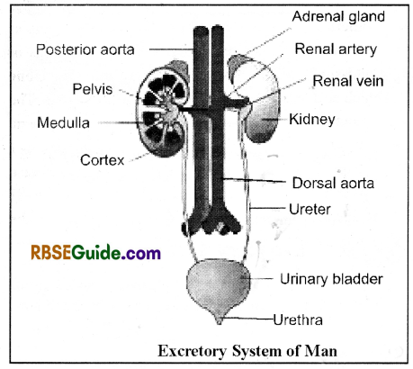
Urinary Bladder
The urinary bladder is baglike which is made up of smooth & involuntary muscles. The lumen of the urinary bladder is lined by transitional epithelium which has great power of stretching. Hence, the urinary bladder can store more urine.
The urinary bladder leads into male urethra & it’s opening is regulated by a sphinctor. In human beings, the sphinctor is made up of voluntary muscles. The sphinctor is relaxed only at the time of micturition.
The act of passing urine is called as micturition which is a partly voluntary process in human beings.
In male, the urethra leads outside through the penis and in female through the vulva.
In woman urethra is absent. Hence, the urinary bladder opens outside directly through urinary orfice.
In man, the urethra can be divided into three parts-
- Prostatic or Urethral Part-Anterior most part having length 2.5 cm. It receives prostate gland, urinary bladder and both the vas deferens.
- Membranous part-It is the middle part which receives the Cowper’s glands.
- Penile part-It is situated inside the corpus spongiosum of penis. It’s length is about 15 cm.
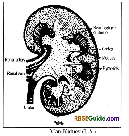
Internal Structure & Histology of Kidney
The kidney is bounded by a renal capsule which is called as tunica fibrosa.
The kidney is divisible into two parts –
- Cortex – Outer part
- Medulla – Inner part
The projections of the medulla into the cortex are called as pyramids. The number of pyramids in the kidney of man is 10 to 12.
The extensions of the cortex into the medulla are called as renal column of Bertini.
In human beings, there are about 20 lakh nephrons in both the kidney.
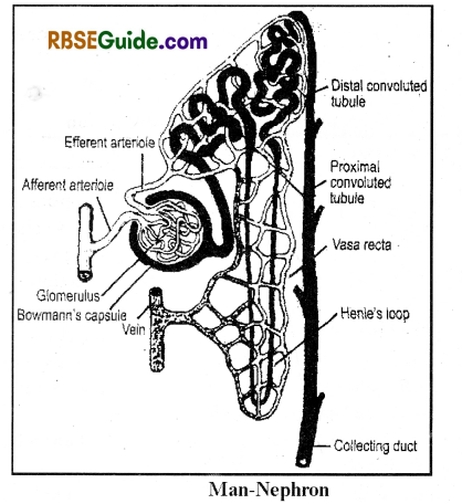
Structure of Nephron or Uriniferous tubule
Structure of Nephron
It is a unit of excretion in which urine is formed independently.
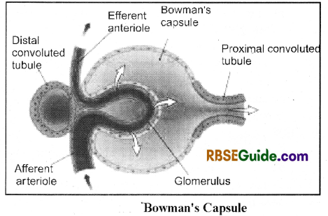
It’s anteriormost part is called as Malpighian body which includes a cup-like Bowmann’s capsule & a glomerulus. The glomerulus is a bunch of blood capallaries.
It receives the blood through a affarent arteriole and the blood comes out through the efferent arteriole. The diameter of the affarent is comparatively more.
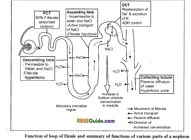
The Bowmann’s capsule opens into a proximal convoluted tubule (PCT) through a short & straight neck. The anterior part of the PCT is more coiled where as its posterior part is almost straight. The PCT opens into a Henle’s loop.
The Henle’s loop is a U-shaped structure which has a distinct descending limb & an ascending limb. The ascending limb opens into the DCT.
The descending convoluted tubule (DCT) is a coiled duct.
Many DCT unite to form a collecting duct. The collecting ducts of one pyramid unite to form a duct of Bellini. The ducts of Bellini lead into the pelvis part.
![]()
The Malpighian body and a part of PCT & DCT are situated in the cortex. Most of the part of PCT & DCT, Henle’s loop and collecting ducts are found in the medulla.
The efferent arteriole forms a peri-tubular capillary network around the PCT, DCT & Henle’s loop which is called as Vasa recta.
The capillaries of Vasa recta join to form venules which . finally open into the renal vein.
Histology of Nephron
The wall of the Bowmann’s capsule is made up of simple squamous epithelium. It’s inner wall has special cells, called podocytes. The podocytes lias finger-like processes which bind the glomerular-blood capallaries.
The neck of the nephron & collecting duct are made up ciliated epithelium.
PCT is made up of simple cuboidal epithelium. It has brush-border of microvilli.
The- Henle’s loop is made up of simple squamous epithelium.
DCT is made up of simple cuboidal epithelium. It is without microvilli.
Types of Nephrons
On the basis of structure, position in the kidney and size of Henle’s loop the nephrons are of two types-
- Cortical nephrons: They are 80% of total nephron. They are found in the outer part of the cortex. Their Henle’s loop is smaller.
- Juxta-medullary nephrons: They are found in the inner region of the cortex. They are 20% of the total nephrons. Their Henle’s loop is longer.
The juxta,medullary nephrons have a juxta glomerular region or juxta-medullary region between the DCT and glomerulus, afferent & efferent arterioles.
It acts as endocrine part. This region has special Lacis cells or Polkisson’s cells. These cells secrete renin hormone which acts on angiotensinogen found in the plasma.
The angiotensinogen is formed in the liver. The renin hormone converts angiotensinogen into angiotensin – I (decapeptide) which by the action of angiotensinase converts into angiotensin-II The angiotensin-II (octapeptide) acts as vaso-constrictor & increases the blood pressure.
The part of DCT forming juxta-medullary region has special macula densa cells in the wall. These cells secrete the bone marrow
Mechanism of Excretion
Urine formation:
Urine forms in the nephrons and according to Cushny, it takes place in 3 steps
- Ultrafiltration
- Reabsorption
- Tubular excretion or Secretion
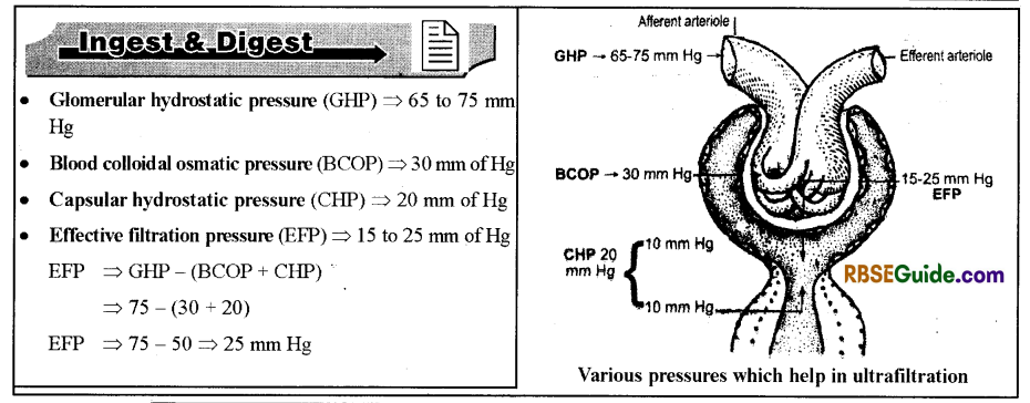
Ultrafiltration
It is a pasive process which takes place from the glomerulus into the RowaniTs Capsule The glomerular epithelium has various micropores (diameter = 0. 1μ.) which increase the rate of filtration.
The ultrafiltration takes place due to increased blood pressure in the glomerulus which is increased due to the difference in the diameters of affarent & efferent arterioles.
There is ultrafiltration of all the componants of the blood except the blood corpuscles, plasma proteins macromolecular foreign substance. The ultrafiltration forms glomerular filtrate in the Bowmann’s capsule.
The glomerular filtrate is a plasma without plasma protein.
Glomerular filtrate => Blood – Blood Cells + Plasma Proteins
Glomerular filtrate => Plasma – Proteins
![]()
The hydrostatic pressure in the glomerulus (GHP) is 65 to 75 mm of Hg. Out of it, the blood colloidal osmotic pressure (BCOP) due to plasma proteins is 30 mm of Hg.
On the contrary, the capsular hydrostatic pressure (CHP) is 20 mm of Hg. Hence, the net Effective glomerular filtration pressure (EFP) is 15 to 25 mm of Hg which is responsible for ultrafiltration.
In human beings, the glomerular filtration rate (GFR) is 125 ml per minute i.e. both the kidneys form 125 ml of filtrate per minute. Hence, 180 litre of filtrate is formed per day. Out of it, only 1.5 litre of urine is produced per day which is 0.8% of the total filtrate.
Reabsorption
In the renal tubules 99.2% of the glomerular filtrate is reabsorbed into the blood.
The reabsorption takes place by active transport and most of the reabosorption takes place in the PCT. However, the water is absorbed by passive transport.
The reabsorption of the substance is according to the renal threshold value. The maximum capacity of both the kidneys to absorb a particular substance is called as renal threshold of the substance.
The renal threshold value of the substances is according to their requirement in the body.
The substances are of 3 types on the basis of their renal threshold value
(a) High threshold substances:
- Such substances are absorbed almost all.
- Example – Sugar, amino acids, vitamins etc.
(b) Low threshold substances:
- They are absorbed in low concentration.
- Example – Urea, creatine, creatinine, phosphate etc.
(c) Athreshold substances:
- They are not absorbed.
- Example – Uric acid etc.
If any substance exceeds its renal threshold value, it will appear in the urine.
The renal threshold value of glucose is 350 mg per minute which is also called as TmG
The reabsorption of the substances is under the control of hormones which are as follows :
(a) Glucocorticoids:
They are secreted by adrenal cortex & they control reabsorption of sugars, amino acids & vitamins in the PCT.
(b) Mineralocorticoids:
They are secreted by the adrenal cortex and they control reabsorption of mineral ions in the PCT.
(c) Calcitonin & Parathormone:
They control absorption of Ca in the DCT.
(d) Vasopressin or ADH:
It is a hormone of Pituitary gland which controls absorption of water in the DCT.
Most of the water (about 80%) is absorhed in the PCT along with the substances. It is called as obligatory’ water reabsorption.
Remaining water (about 20° o) is absorbed in the DCT under the control of ADH according to the body requirement. It is called as facultative water reabsorption.
Excess release of the ADH result in decreased volume of the urine.
![]()
Tubular excretion or Secretion
It is an active process which takes place in the comers of the Henle’s loop. The cells of these corners take inacromolecular waste products from the blood of vasa recta & excrete them into the lumen of the renal tubule. Normally urea, medicines, uric acid, hipuric acid etc. are excreted by this process.
Osmolarity of filtrate in Nephron
The osmolarity of the filtrate in the Bowmann’s capsule & PCT remains unchanged i.e. it is isotonic to the blood plasm.
The descending limb of the Henle’s loop is permeable to the water but impermeable to the solutes. Hence this part absorbs only water. Therefore the filtrate becomes Hypertonic.
The permeability of the asceding limb of the Henle’s loop is changeable. Normally, it is impermeable to water but permeable to NaCI. Hence, Na++ are reabsorbed in this part making the filtrate hypotonic.
In DCT the glomerular filtrate is hypotonic.
The upper part of the collecting duct is permeable to water & the basal part is permeable to urea. Hence, due. to reabsorption of water, the glomerular filtrate becomes hypertonic. In the last part of the collecting duct, the filtrate is called as urine.
The osmolarity sequence in the urine formation is –
Isotonic → Hypotonic → Hypertonic
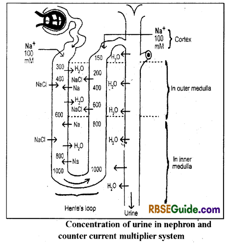
Mechanism of urine concentration
Mammals form hypertonic urine The urine is made hypertonic with the help of counter current multiplier system. This process takes place in the Henle’s loop and It involves mainly Na+ & CE
There is osmotic withdrawal of water from the collecting duct to make the urine hypertonic.
The ADH makes the wall of collecting duct more premeable to the water.
The descending limb of the Henle’s loop is permeable to water. It’s surrounding tissue fluid is hypertonic. Hence, the water moves out & the Na+ & Cl– move in the descending limb by passive transport. Therefore, the filtrate in the descending limb finally becomes hypertonic.
The ascending limb of the Henle’s loop is impermeable to the water. The Na+ & Cl– move out by active transport. Hence, the filtrate finally becomes hypotonic. The Na+ & Cl– re-enter into the descending limb of the Henle’s loop.
The collecting duct always passes through the hypertonic tissue fluid. Hence, water comes out osmotically making the filtrate hypertonic.
Other Excretory Organs in Man
(1) Skin : Sweat glands are found in the human skin, which secrete out sweat containiang water and some nitrogenous excretory substances.
(2) Lungs : Cellular respiration release C02 as excretory substance, which is eliminated outside by respiratory process in the lungs.
(3) Liver : Liver cells convert nitrogenous part of amino acids into ammonia and then ammonia into urea. The urea is released into the blood. The liver also forms bile pigments which are excreted along with the bile juice into the intestine.
Disorders related to Excretion
Irregularities in excretion may cause various diseases in human. Some of the diseases are as follows:
(1) Uremia : When amount of urea in the blood becomes more than 10-30 mg/100 ml, the condition is called Uremia. Excess amount of urea in blood is harmful and may be fatal.
![]()
(2) Gout: It is hereditary disease in which blood uric acid is increased. It get deposited in the synovial joints and in Kidney tissues. This disease may cause due to dehydration, fasting and diuresis.
(3) Kidney stones : Normally crystals of substance like calcium oxalate, phosphate, uric acid etc get deposited in the renal pelvis as renal stones. They cause pain & dificulty in micturition.
(4) Bright’s disease or Nephritis : This disease is caused due to infection of Streptococci bacteria in the glomeruli. As a result, they swells and their membrane becomes more permeable to RBC’s and proteins which filtered out and appears in the filtrate. If nephritis is not cured than fluid deposition occurs and condition of swollen legs happens, which is called Edema or Dropsy.
(5) Glycosuria : Presence or excretion of glucose in the urines is called Glycosuria. It is caused due to deficiency of insulin hormone. This disease is called as diabetes mellitus.
(6) Disuria : Pain at the time of micturition is called disuria.
(7) Polyurea : The increase amount of urine due to less absorption of water is called as polyurea.
(8) Cystitis : The swelling of urinary bladder by the infection of bacteria, chemical or mechanical damage is called cystitis.
(9) Diabetes Insipidus : Due to hyposecretion of antidiuretic hormone (ADH), the water absorption does not occur in the distal convoluted tubuler and collecting tubule results into increase in the volume of Urine. There is frequent and excess micturition.
(10) Oligourea : Formation of less quantity of urine as compared to normal.
(11) Proteinurea : Presence of more protein in urine is called proteinurea.
(12) Albuminurea : Presence of more amount of albumin protein in the Urine.
(13) Ketonurea : Increase amount of ketone bodies in urine such as acetone acetic acid.
(14) Haematonurea : Elimination of red blood corpuscles (RBC’s) along with urine is called haematonurea.
(15) Haemoglobuinurea: Presence of haemoglobin in urine is called haemoglobinurea.
(16) Pyurea: Presence of pus cells in urine is called pyurea.
(17) Jaundice : Presence of bile pigments in huge amount in urine is a symptom of jaundice. It is seen during hepatitis or due to blockage of bile duct.
(18) Alkaptonurea: Presence of alkaptone or homogentisic acid is termed alkaptonurea. When alkaptone comes in contact with air than urine becomes black colour, it is also known as Black urine disease.
Haemodialysis and Artificial Kidney
Malfunctioning of Kidneys causes increased amount of urea into the blood. It is termed as uraemia.
The process by which waste products are isolated from the blood is called as haemodialysis.In this process blood is removed slowly from a major artery and transfer it to a machine called a dialyser or dialysis machine.
It is also called as artificial kidney. Blood temperature lowered upto 0°C and an anticoagulant heparin is added into the blood.
The dialysis machine is made up of a series of cellophane membranes that act as filter and a special liquid called dialysate (dialysis fluid). The membrane filters waste products including urea – pass into the dialysate fluid.
These membranes are impermeable for large protein molecules and permeable for urea, uric acid, creatinine and mineral ions.
The process of separating smaller molecules from larger molecules that occurs in the artificial kidney, is called as haemodialysis.
The blood again brought at body temperature by slightly heating it and mix antiheparin to neutralize the heparin which was earlier mixed.
This purified blood slowly enter into the body via a major vein. It is necessary for a person in which kidneys are not functioning to enhance life.
![]()
Kidney Transplantation
In human beings when kidneys stop functioning and are not curable than a kidney of other healthy person is to be transplant. The Kidney is obtained from a donor who may be decreased or live.
Kidney donor should be nearest relative whose blood and tissue structures are to be similar.
If unmatched kidney transplantation is done than the immune system of patient will not accept the transplanted kidney and it will not work properly so patient may die.
Although special medicines are given to patient to inactivate immune system and possibility to accept kidney of donor is increased.