
RBSE Class 12 Biology Notes Chapter 27 Man-Sensory Organs (Sense Organs)
General:
There are many sense organs in the body to receive various stimuli.
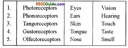
Sherington classified the sense organs into three groups:
(a) Exteroceptors:
Example-Tangoreceptors, Phonoreceptors, Rheoreceptors, Algisoreceptors, Skin receptors etc.
(b) Interoceptors:
Example-Sense organs found in the visceral organs
(c) Propioceptors:
Example-Sense organs found in voluntary muscles, tendons, joints, ligamens etc.
Parker classified the sense organs into 3 groups on the basis of nature of stimuli-
(a) Chemoreceptors:
Example-Gustoreceptors, Olfactoreceptors
(b) Mechanoreceptors:
Example-Algisoreceptors,Tangoreceptors, Pressuroreceptors etc.
(c) Radioreceptors:
Example-Thermoreceptors, Photoreceptors etc.
Both the extero & intero receptors remain interconnected by somatic & visceral sensory nerve fibres. .
The receptors send the stimuli to the central nervous system.
Following sense organs are found in human beings-
![]()
General
There is a pair of eyes in rabbit which are situated on both the lateral sides of the head in the eye orbits movably.
Each eye remains attached with the help of 6 eye muscles.
In man, 2/3 part of the eye ball is inside the eye orbit & 1/3 part remains outside.
The exposed part of the eye ball is covered by a modified skin which is called as conjunctiva. It is transparent and it is modified epidermis.
The exposed part of the eye is gaurded by a pair of eyelids viz.-upper & lower, eye-lids.
The margins of the eye-lids are provided with hair which are called as eye lashes.
Each eye is covered by nictitating membrane which is reduced in rabbit and functional in hare.
In human beings, vestigeal nictitating membrane is found in the inner comer of the eye.
Muscles of Eye Orbits
Each eye is attached movabally with the help of 6 eye muscles in the eye orbit.
These eye muscles are voluntary & striated.
There are 4 rectus & 2 oblique eye muscles which are as follows:
1. Anterior or external rectus. (Lateral Rectus):
- It is attached to the outer lateral side of the eye ball
- It receives Abducens cranial nerve.
2. Posterior or internal rectus. (Medial Rectus):
- It is attached to the inner (Medial) side of the eye ball.
- Occulomotor cranial nerve innervates it.
3. Superior rectus:
- It is attached to the mid-dorsal side of the eye ball
- It receives occulomotor cranial nerve.
4. Inferior rectus:
- It is attached to the midventral side of the eye ball
- It receives oculomotor cranial nerve
5. Superior oblique muscle:
- It is attached between the external rectus & superior rectus.
- It receives trochlear cranial nerve.
6. Inferior oblique muscle:
- It is attached between the inferior rectus and external rectus.
- It receives occulomotor cranial nerve.
Anterior rectus & posterior rectus muscles rotate the eye ball inside & outside.
Superior rectus & inferior rectus rotate the eye balls upside & downside.
![]()
Internal Structure of Eye
Wall of Eye
The wall of the eye consists of following Layers-
(a) Sclerotic:
- It is the outermost layer of the sclerotic which gives shape to the eye & protects it.
- It is made up of thick fibrous connective tissues.
- Its outer part modifies to form cornea. The cornea focuses the light rays on the lens.
- In eye, only cornea part can be transplanted.
(b) Choroid:
It is the middle layer of the eye ball which is vascular.
It has branched pigmented cells. In rabbit, its color is red.
Its outer part modifies to form a diaphragm-like iris. The iris has a central pore called pupil.
The pupil is surrounded by a sphinctor which is made up of smooth muscles. It controls the diameter of the pupil. The iris also has radial muscles which dilates the pupil.
The iris functions like a diaphragm of a camera.
The iris muscles are ectodermal in origin.
The choroid layer forms a folded ciliary body at the base of the iris. The ciliary body is a highly sensitive structure. The ciliary body has small ciliary processes & their number is 70 to 80 in human beings.
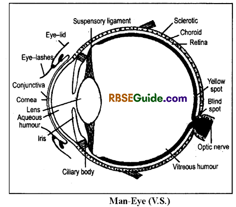
(c) Retina:
It is the innermost layer of the eye ball which is photosensitive.
It has four layers
(i) Pigmented layer
- It is the outermost layer which has a layer of melanin-containing hexagonal cells.
- It forms numberous microvilli on the inner side.
- It provides support to the retina.
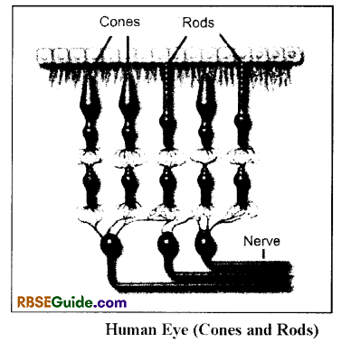
(ii) Layer of rods & cones
This layer is made up of rods & cones. Their number is different in different species & they are sensory to light.
The rods are long & cylindrical cells which are functional even in dim light i.e. they help in vision both in night and in dim-light.
The cones are small & club-shaped which are functional in moderate to bright light. They are responsible for colour vision.
The axis passing through the centre of cornea & lens is called as optical axis.
There is visual axis just above the optical axis.
The part of retina at the visual axis is called as yellow spot or macula lutea. The centre of the yellow spot is called as fovea centralis. The yellow spot is made up of only cones & it is the site of clearest vision.
The margins of the retina are called as orra serrata which has less number of rods & cones.
The point of origin of optic nerve from the retina is called as blind spot. It is without rods & cones.
The cones are of three types viz.-red cones, green cones & blue cones.
![]()
(iii) Layer of bipolar neurones
- It consists of bipolar neurones.
- Their dendrites form synapses with the processes of rods & cones which is called as outer plexiform layer.
(iv) Ganglionic layer
- It also contains bipolar neurones.
- Their dendrites form synapses with the axons of the layer of bipolar neurones. It is called as inner plexiform layer.
- Their axons are long & they form an optic nerve.
- the optic nerve comes out of the wall through an optic for a man.
Lens
It is a biconvex & transparent structure which is situated just behind the pupil.
The lens is attached to the ciliary body with the help of suspensory ligament. The suspensory ligament is also called as zonula of Zinn.
The lens is situated in a transparent capsule.
The shape of the lens is changeable which are with the help of ciliary body.
The convexity of the lens increases to see the near objects and the convexity decreases to see the far objects. It is called as eye accomodation.
The lens focuses light rays on the retina.
Intraocular Cavity
The eye ball has an introoceular cavity which is divisible into an anterior aqueous chamber & a posterior vitreous chamber.
The aqueous chamber is found in between cornea & lens. It is full of aqueous humour w hich is secreted by ciliary body.
The aqueous humour consists of water, O2, CO2, glucose & other nutrients. This fluid provides nutrients to the eye.
The vitreous chamber is found in between the lens & retina. It is full of vitreous humour. It is also secreted by ciliary body. It has about 90% water, vitrein mucoprotein, hyaluronic acid & some salts. The vitreous humour also contains small & thin collagen fibres & hyalocytes.
The refractive index of the aqueous humour is similar to cornea & of vitreous humour is similar to the lens.
![]()
Eye Glands
Three types of glands are found in the eyes-
(i) Meibomian glands:
- These glands are found at the innersides of the eyelids.
- They secrete an oily substance which lubricate the eye lids and facilitate movements of the eye lid.
(ii) Glands of Moll’s:
- They are also called ciliary, glands.
- They are modified sweat glands.
- They open at the margins of eyelids near the follicles of eye lashes
(iii) Lacrymal Glands:
- There are three tear glands in each human eye which are also called as Lacrymal glands.
- They secrete a watery fluid which keeps the eyes moist and cleans the eyes. This fluid comes out in the form of tears when secreted in excess.
- The tears are salty in taste and contain Lysozyme which acts as antibacterial.
- Normally, the watery fluid (tear) drains into the Nasal cavities through minute Naso-Lacrymal ducts. It comes out in the form of tear.
Working of Eye
The eye functions like a camera. It’s working involves following events
Light communication:
The light rays reflected by an object enter into the eye after crossing through transparent structures such as conjuctiva. cornea aqueous numous lens and vitreous humour.
Formation of Image:
The lights rays which entered into the eye, finally forms an image on the retina.
Type of Image:
The image of object forms in the human eyes is always real but inverted.
Formation and sending of Nerve impulse:
The rods & cones found in retina generate a nerve impulse by converting light energy into chemical energy. Such nerve impulses are transmitted to the brain through the optic nerve.
Analysis of Impulses:
The brain analyses the impulses of the image, and identify the straight image. Though, the image forms on retina is inverted.
For Farsightedness:
The lens focuses the light rays on the retina to form an image. For distant vision, the lens convexity decreases to focus the rays on the retina.
For Shortsightedness:
The lens convexity increases to see the near objects. It is with the help of ciliary’ bodies.
The capacity of human eyes to see the objects of vivid distances is called as eye accommodation.
![]()
Binocular vision:
In all primates including man, both the eyes are closely located in front. In this arrangement, images can overlap and give feelings of depthness which is known as 3-D vision or binocular vision.
Biochemistry of Vision
Formation of image in the eye involves many types of biochemical changes.
The retina consists of rods & cones.
The rods are responsible for general vision. These rods consist of visual pigment called rliodopsin or visual purple.
It changes in the presence of light into Leumorliodopsin. Leumorhodopsin changed into metarhodopsin which finally convert into a protein scotopsin and retinin. In darkness again setopsin and retinin unite to form rhodopsin.
Retinin is a derivatives of vitamin-A. Hence for the healthy eyes vitamin-A is essential. In the absence of Vitamin-A there is no formation of rhodopsin which create night blindness in which person unable to sec in dim light.
Another pigment iodopsin is found in the cones. Which is responsible to know about colour differentiate. Three kinds of cones are present to differentiate various colours such as red, green and blue cones. Amongst these for red colour is erythrolabe, green colour is chlorolabe and for blue colour is cyanolabe pigments are found.
Common Eye Diseases
The various eye diseases are as follows-
Myopia:
- It is an eye disease which may occur to any person at any age.
- The person is unable to see distant objects clearly.
- It is caused either due increased focal power of the lens (lens become more convex) or eye ball becomes larger.
- It is also called as short sightedness.
- It can be corrected by used of biconcave lenses, which are also called as minus lenses.
Hypermetropia:
- It is also called as far sightedness.
- The person is unable to see near objects clearly.
- It is caused either reduced focal power of the lens (lens becomes less convex) or the eye ball becomes smaller.
- It can be corrected by the uses of biconvex lenses which are also called as plus lenses.
Catarac:
- It is an eye disease of old age.
- The eye lens gradually becomes opaque, as a result the light rays fails to enter into the eye.
- Cataract disease is also known as safaid motia.
- Surgical removal of lens and either implantation of artificial lens or use of spectacles with convex lens is a corrective measure.
Astigmatism:
- It is due to abnormal shape of cornea, in which different curvature in different regions of cornea appears.
- It can be corrected by using cylindrical lenses.
- It can be corrected by the use of cylendrical lenses.
Conjuctivites:
- This eye disease is caused due to viral or bacterial infection.
- There is inflammation & irritation in conjuctiva. The conjuctiva also becomes red.
![]()
Colourblindness
It is a hereditary eye disease.
It was first described by Horner. It is of 2 types
- Red-green colourblindness
- Blue colourblindness
The red-green colourblindness is most common & it is also called as proton defect. Red – green colourblindness is caused due to absence of red/green cones in the retina.
The person is unable to identify red, green, yellow & orange colours. These colours appear to be green in absence of red cones and they appear to be red in absence of green cones.
It is a sex-linked disease which is caused due to a recessive gene on the x – chromosome. The mother (female) acts as a carrier.The father of colourblind daughter is always colourblind.
In the F1 generation of carrier mother & normal father, 50% generation will be normal, 25% will be carrier & 25% will be colourblind.
In F1 generation of carrier mother and colourblind father, 50% daughters will be colourblind & 50% will be carrier likely, 50% boys will be normal & 50% will be colourblind.
In the F1 generation of normal mother & colourblind father, all males will be normal and all daughters will be carrier.
Ear – Hearing & Balancing Organ
general
- There is a pair of ears in mammals on both the sides of the head.
- It is situated in an auditory capsule which consists of a periotic bone & tympanic bull bone.
- The ear acts as stato-acoustic organ i.e. sensory to sound & body balance.
- The mammalian ear has three parts viz.- external ear, middle ear & internal ear.
External Ear
This part includes pinna & external auditory meatus.
The pinna in rabbit is long, movable & hairy.
The pinna in man in vestigeal & immovable.
The pinna is made up of fibrous elastic cartilage. It sends the sound waves into the external auditory meatus.
The external auditory meatus is a samll tube-like structure & its length is about 2.5 cm. The external auditory meatus has many ceruminous glands which secrete ear wax or cerumin. The cerumin makes the tympanum water proof. The external auditory meatus also provided with few hair.
The external auditory meatus of some man has long &, dense hair. It is called as hypertrichosis. It is a Y-Iinked hereditary disease.
There is a tympanum or ear drum at the end of the external auditory meatus which is attached to the periotice bone with the help of a cartilage called annulus tympanicus.
![]()
Middle Ear
It is found in the tympanic bulla & also called as auditory cavity.
The middle ear opens into a pharynx through a eustachian tube.
There are 2 windows between the middle & internal ears
(i) Fenestra ovalis
- It is oral in shape & it is covered by a thin membrane.
- It remains connected to the scala vestibula of the internal ear.
(ii) Fenestra rotundus
- It is round in shape & it is covered by a thin membrane
- It remains associated to the scala tym-pani of the internal ear.
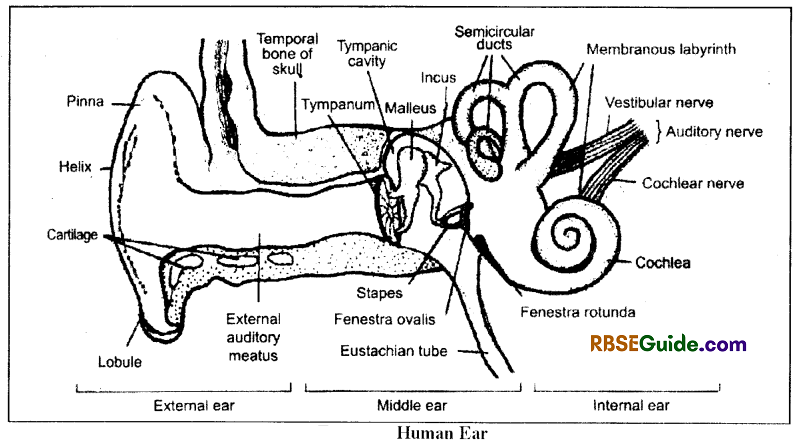
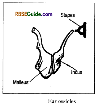
- The middle ear has three smaller ear ossicles or bones which are movable attached between the tympanum & the fenestra ovalis. The ear ossicles remain attached with the help of elastic ligaments & are as follows-
(i) Malleus
- It is largest & hammer-shaped. It is formed by the modification of articular bone.
- It is attached to the tympanum by tensor tympanic muscles.
- It is attached to the incus bone by synovial hinge joint.
(ii) Incus
- It is anvil or club shaped and it is formed by the modification of quadrati bone.
- Its inner end remains attached to the stapes bone with the help of synovial ball & socket joint.
(iii) Stapes
- It is the smallest bone of the body which is stirrup
- The tympanum separates external & middle ears.
- Its weight in man is 1.2 mg.
- It remains attached to the membrane of fenestra oralis.
- It is a modified hyomendibular bone.
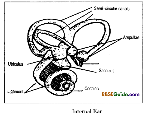
Internal Ear
It is a semi-transparent structure which is also called as membranous labyrinth. It is situated in a bony labyrinth.
The internal ear is surrounded by a perilymph.
It has 2 types of parts
Vestibule
It has three semicircular canals which originates from utriculus at 90°. They are called as anterior, posterior & external semi-circular canals.
The anterior posterior semi-circular canals have a common origin which is called as crus commune.
The distal end of the semi-circular canal forms an ampulla. The ampullae of anterior & external semicircular canals remain closely situated.
The seme-circular canals & the ampullae are filled with a viscous fluid called endolvmph.
Each ampulla has a sensory crista. The crista is lined by a sensory epithelium provided with stereocilia & kinocilia. The kinociliuin is a movable flagellum.
The crista has an otoconia or otolith in the centre which is made up of CaCO3 The otoconia moves here & there in the endolymph. The cristae are sensory to the dynamic balance.
There is a sacculus near the utriculus which remain connected by a duct. Both the utriculus & sacculus has 1-1 maculla which are sensory to static balance.
The sacculus gives out an-endolymphaticus duct. It remains closed in the mammals. In fishes it opens outside the body.
![]()
Cochlea
It is a spiral tube which originates from the sacculus. Both remian connected by a small duct called ductus reunions.
The cochlea has 2.5 coils in rabbit and 2.75 coils in man. The coils remain linked by soft ligament.
Internally, the cochlea has 3 chambers viz. scala vestibule, scala media and scala tynipani.
There is a basilar membrane between scala media & scala tympani. Similarly, there is a Reissner’s membrane between scala vestibule & scala media.
The scala vestibuli & scala tympani remain united by a duct called helicotrema & they are full of perilymph fluid.
The scala media is full of endolymph fluid.
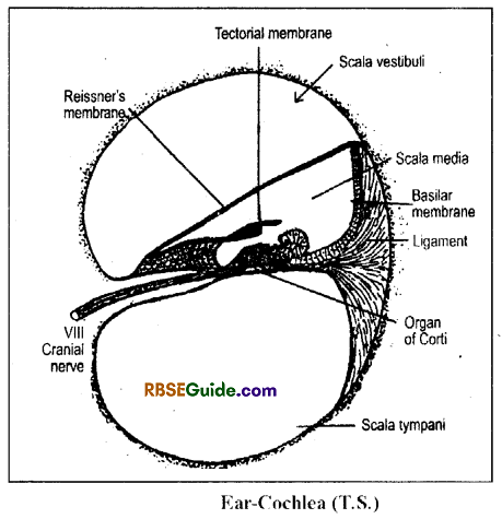
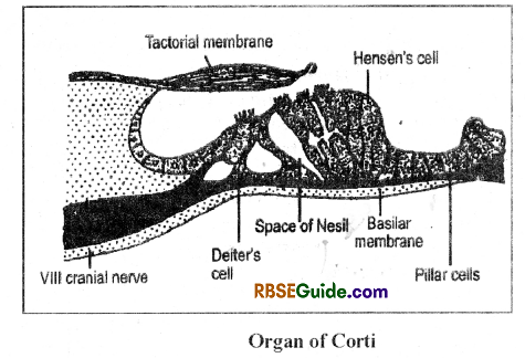
The basilar membrane modifies to form an organ of Corti in the scala media. The organ of corti has three types of cells viz.–Deiter’s cells., Hensen’s cells & pillar cells, these cells are provided with stereocilia & their nerve fibres form VIII cranial nerve.
There is a factorial membrane just above the organ of Corti in the scala media which is formed by the modification of the Reissner’s membrane.
The organ of corti is sensory to hearing.
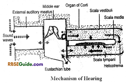
Machanism of hearing:
The pinna collects sound waves & transmit the waves into the external auditory meatus. These waves strike to the tympanum, as a result the tympanum begins to vibrate.
The air imbalance caused in the middle ear due to vibrating tympanum is balanced bv the air displaced through the eustachian tube
The ear ossicles convey the sound vibrations to the fenerstral ovalis. They amplify the vibrations by 2ft times.
The pressure imbalance caused by the vibrating fenestra ovalis membrane is balanced by the membrane of fenestra rotundus
The vibrating membranes of the fenestra ovalis & fenestra rotundus cause the perilymph to vibrate which intum makes the Reissner’s membrane to vibrate.
The vibrating Reissner’s membrane causes the endolymph in the scala media to move. As a result, the factorial membrane moves & strikes to the organ of Coti. Hence, the sound waves reach to the organ of Corti in the form of mechanical stimuli.
The organ of Corti converts these mechanical stimuli into electro-magnetic waves and transmits these waves to the cerebellum through the VIII cranial nerve.
Body balance
Sensory cells or balance receptors are present in the utriculus, sacculus and ampullae of the semicircular canals. These receptors contain the sensory hair which are very sensitive lo the position of head with respect to gravity.
Balance at the time of movement: When body is in motion it perceive by utriculus and saceulus. When a body is in motion, the sensory granules of calcium carbonate called otolith present in endolymph press the sensory hairs, which in turn send impulses to nerves.
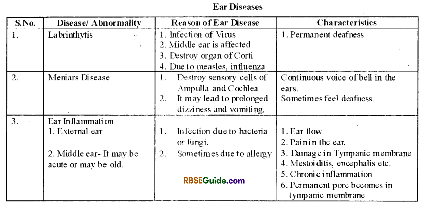
Ear Diseases
Some of the ear diseases are as follows
![]()
Tongue & Nose
Tongue gives the sense of taste and nose gives sense of smell. Both these are chemical senses and they depend on the nature of chemical substance.
Sense of taste is perceived by chemoreceptors present on the tongue. The human tongue has about 10.000 taste buds present on the top and sides of the tongue.
Odours are detected by olfactory hairs (sensory hair) that emerge from the receptors. When molecules of chemicals enter to nose with air. they stimulate sensory’ epithelium of nose, which send impulses to the olfactory region of brain.
Skin
Skinhas terminal ends of various types of nerves. Feeling of various kinds of touch is experienced by these nerves. Some nerves are capable to observe very light touch, some nerves related with high pressure and others for cold, temperature and pain etc.
Sense of hunger is by receptors present in the wall of stomach. Stimulation of nerves present in oesophagus for thirst. Tiredness is experienced by the sense observed by muscles.