Rajasthan Board RBSE Class 12 Biology Notes Chapter 31 Man-Gametogenesis
General
Sexual reproduction involves types of special cells which are called as gametes. These gametes are formed in the gonads from the germ cells and the process of their formation is called as gametogenesis.
Gametogenesis is the primary phase of embryogenesis.
The gametes are of two types viz.-sperms & ova. The sperms are formed in the male gonads, the testes and the ova are formed in the female gonads, the ovaries.
The germ cells in the gonads originate from the primor-dial germ cells which are extra-gonadal in origin
ie. they are formed outside the gonads. After formation, they migrate into the developing gonads and get established to form the germ cells.
In mammals, the primordial germ cells are formed near the yolk sac from the mesoderm.
In reptiles & birds, the primordial germ cells originate from the endoderm which after formation migrate into the gonads.
In amphibians, the primordial germ cells originate from the lateral mesoderm. Later on, they migrate into the gonads through the posterior mesentry.
![]()
Gametogenesis is of two types:
- Spermatogenesis
- Oogenesis
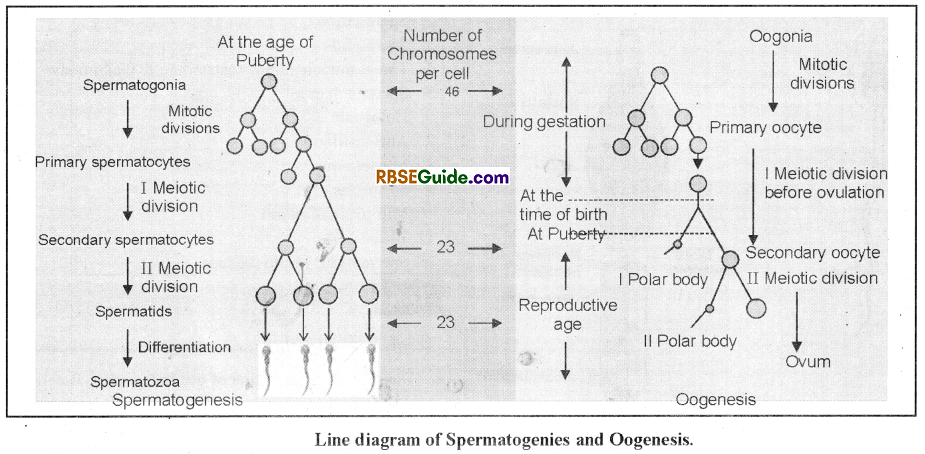
Spermatogenesis
The formation of the sperms from the germ cells in the testes is called as spermatogenesis.
The structural & functional unit of testes is seminiferous tubule. It is provided with germinal epithelium which is made up of germ cells.
Spermatogenesis is a continuous process but for the convenience of study it is divided into two steps
(A) Spermatocytogenesis &
(B) Spermiogenesis
Spermiogenesis
It results in the formation of four haploid spermatids from one diploid germ cell.
It is further divided into three phases :
(a) Multiplication phase
(b) Growth phase
(c) Maturation phase
Multiplication phase
- During this phase the germ cells divide repeatedly by mitosis.
- It results in the formation of more cells which are called as spermatogonia.
- The spermatogonia are diploid.
![]()
Growth phase
- This phase results in the formation of primary spermatocyte.
- It is a phase of short duration which does not involve any division.
- The spermatogonia exhibit growth due to accumulation of proteins & chromatin substances.
The primary spermatocytes are also diploid.
Maturation phase
During this phase, one diploid primary spermatocyte divides meiotically to form four haploid spermatids. It is further divided into 2 phases
(i) Maturation-I
It involves meiosis-I which is also called as reductional division.
The diploid primary spermatocyte divides to form two haploid secondary spermatocytes.
(ii) Maturation-II
It involves meiosis-II which is also called as equational division.
The two secondary spermatocytes divide to form four spermatids.
Spermiogenesis
- During this phase a non-motile & tail-less spermatid transforms into a motile & tailed sperm.
- There is no division during the spermiogenesis and one spermatid forms only one sperm.
- It is also called as spermatoleosis.
- The changes during the spermiogenesis result in the formation of head, middle piece & tail in the sperm.
- The various changes during spermiogenesis are of 2 tvpes-
![]()
Changes in Cytoplasm
During spermiogenesis most of the cytoplasm of the spermatid is l«j»t in the form of cytoplasmic residue.
The other changes in the cytoplasm are as follows-
(i) Changes in the Centriole
- The spermatid has two centrioles. During the process of spermiogenesis, one centriole moves anteriorly and occupies a position in the notch of the nucleus. It is called as proximal centriole.
- The second centriole assumes slightly posterior position and form the neck of the sperm. It is called as distal centriole.
- Both the centrioles in the sperm are situated at right angle to each other.
- The distal centriole grves rise to an axial filament which is found in the middle piece & tail of the sperm.
- The axial filament exhibits 9+2 fibrilar arrangement out of them 9 fibres are peripheral & double while the 2 fibres are central & single.
- In the sperms of some species, an additional ring centriole is present in the posterior part of the middle piece. However, it’s function is obscure.
(ii) Changes in Mitochondria
- During the process of spermiogenesis, all the mitochondria of the spermatid fuse to form a nebenkern or Jensen’s sheath.
- The nebenkern along with little cytoplasm form a spiral sheath around the anterior end of the axial filament which is called as manchette.
- The nebenkern provides required energy to the spenn.
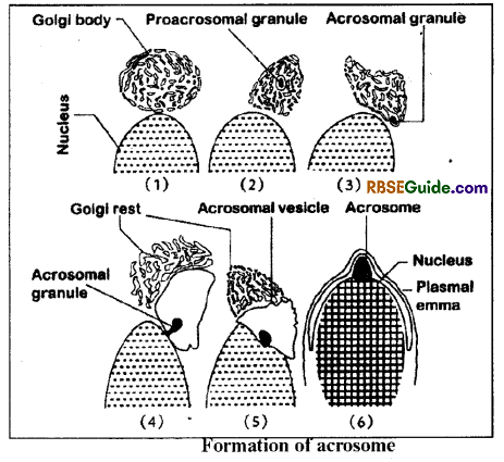
(iii) Formation of Acrosome
- The anteriormost part of the sperm is called as acrosome. It is formed by the Golgi body during the spermiogenesis.
- Firstly, one of the vacuole of the Golgi body develops an acrosomal granule. Gradually, the acrosomal granule accumulates lytic enzymes and forms a cap-like structure on the anterior end of the nucleus.
- Soon this cap get bounded by a single unit plasma membrane and forms an acrosome.
- The rest parts of the Golgi body are lost in the form of Golgi rest.
![]()
Changes in the Nucleus
- There is loss of water and unwanted substances from the nucleus during spermiogenesis.
- The nucleolous & RNA are lost gradually.
- The nucleus slightly elongates and become narrow.
- The remaining DNA with nucleoproteins get condensed in the nucleus.
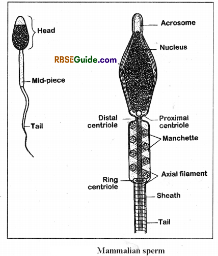
Structure of Human Sperm
- Male gamete is called as spermatozoon or sperm. It is as single cell structure and it bear a haploid set of chromosomes in the nucleus.
- A human sperm has a head, neck, mid – piece & tail.
Head
- It is the anterior part of the sperm. It’s shape varies from species to species.
- The anteriormost part of the head is occupied by the acrosome. The aerosome is formed by the Golgi-body and it is bounded by a unit membrane. It is full of sperm lysins & it helps in fertilization.
- There is a nucleus just behind the acrosome which is slightly elongated and it’s posterior end has a notch. The nucleus has’a haploid set of chromosomes which have protamines in addition to DNA.
- The sperms of some species have a vacuole-like structure between the acrosome and nucleus which is called as perforatorium.
- The posterior notch of the nucleus harbours a proximal centriole which is to be contributed to the egg at the time of fertilization. It helps in the begining of first cleavage.
Neck
- It is a small part which connects head to the mid-piece. It. bears a distal centriole which remains at 90° to the proximal centriole.
- The distal centriole gives rise to an axial filament which extends through out the length of mid-piece & tail. It exhibits a fibrilar arrangement of 9 (doublet) + 2 (singlet).
![]()
Middle piece
- In the middle piece, the axial filament is surrounded by sheath called manchett. The manchett is made ‘up of nebenkern (fused mitochondria) and little condensed cytoplasm.
- In the sperms of some species, the posterior end of the middle piece may has a ring centriole. It’s role is not clear.
Tail
- In the tail, the axial filament is covered by a sheath which is made up of 9 (singlet) fibres. This sheath is absent arround the terminal end of the tail.
- The whole sperm is bounded by a plasmalemma.
- Most of the sperms are mono-flagellated.
Oogenesis
General
The process of formation of ova in the ovary is called as oogenesis.
The germ cells are found in the form of germinal epithelium around the ovary.
The process of oogenesis is a continuous process but for the convenience of study it is divided into three phases:
(A) Multiplication phase
(B) Growth phase
(C) Maturation Phase
![]()
Multiplication
- During this phase, the germ cells divide reapedly to form oogonia.
- The oogonia are diploid and are also called as egg mother cells.
Growth phase
- It is the longest phase of the oogenesis which does not involve any division.
- The oogonia during growth phase enlarge to form primary oocyte which are diploid.
- The growth in the oogonia is different in different species. For example, it is 20-times in mammals, 200-times in amphibians and 4000- times in birds.
- The growth rate of oogonia varies from species to species. For example, in woman it’s duration is of 14 days, in then the growth takes place in 6 to 14 days and in frog it is of 3 years.
- In frog, the ovary lacks mature ova in the first two years.
- The growth phase is further divided into 2 sub-phases viz.-
(a) Pre-vitellogenesis and
(b) Vitellogenesis
Pre-vitellogenesis
This sub-phase involves following 2 types of changes-
(i) Changes in the Ooplasm
The cytoplasm of the oogonium is called as ooplasm. During this phase the ooplasm becomes more granular & increases in amount.
The mitochondria increase in number and they form groups which are called as mitochondrial clouds. Because of increased mitochondria the amount of mDNA becomes more than the nuclear DNA (Epel, 1973). The mitochondrial clouds are also called as yolk nuclei of Balbiani.
The Golgi body becomes hyperactive & secretes cortical granules. These granules are full of mucopoly-saccharides and get arranged in the peripheral part of the ooplasm. At the time of fertilization, these cortical granules form the fertilization membrane. The cortical granules are absent in the eggs of rats, insects, birds & guinea pig.
The endoplasmic reticulum is found mainly in the form of annulated lamellae & small vesicles. The annulated lamellae store RNA. According to De Robertis, these lamellae get disintegrated at the end of oogenesis.
(ii) Changes in the Nucleus
The size of the nucleus is increased gradually due to increased amount of nucleoplasm.
The nucleoli increase in size & particularly in amphibians, the number is increased upto 600 to 1200.
Formation of more amount of mRNA, tRNA and rRNA from the nucleolar organizer parts of the chromosome. It is called as gene amplification by Epel (1973). De Robertis (1975) termed it as redundancy.
Some of the mRNA in the cytoplasm form informosomes (Spirin, 1965). Each informosome has a molecule of mRNA bounded by a protein layer. These informosomes are used in protein synthesis during emergency.
The nucleolous stores the proteins & RNA which are required in the formation of ribosomes.
In some fishes, amphibians and reptiles, the nucleus develops Lamp-brush chro-mosomes during the growth phase to form large amount of mRNA.
![]()
Vitellogenesis
This sub-phase mainly involves formation & storage of yolk or vitellin. The yolk is a stored form of food which consists of proteins, phospholipids & neutral fats.
The formation of yolk is extra-gonadal i.e. it takes place outside the ovary. In insects the vitellogenesis takes place in the fat body and in the chordates, it occurs in the liver.
The yolk form is transported insoluble into the ovarian cells by the help of blood. The Golgi body & ER transfer the soluble yolk into the mitochondria clouds which convert it into insoluble form. Hence, the mitochondrial clouds help in the storage of the yolk.
The yolk of hen’s egg contains 32 to 33% phospholipids, 16 to 17% proteins (albumin, phosvitin, lipovitellin), 10% carbohydrates & 40-42% water.
The yolk is of two types-
- Protein yolk – It has more amount of proteins.
- Fatty yolk – It has more amount of fats.
The yolk is found in 2 forms:
(i) Granular yolk
- It is found in the form of small granules.
- It remains uniformly distributed in the ooplasm.
- Eaxmple : Protochordates.
(ii) Yolk platelets
- In most of the chordates, the yolk is found in the form of large granules which are called as yolk platelets.
- In amphibians, the yolk platelets are oval & flat. The platelets are made up of phosvitin & lipovitellin.
- Phosvitin has 8.4% proteins whereas the amount of protein in lipovitellin is 17.5%.
- Each yolk platelet has a molecule of. lipovitellin & two molecules of phosvitin.
Maturation phase
The maturation phase of oogenesis involves meiosis. It finally results in the formation of one haploid ootid & three haploid plocytes or polar bodies.
It is further divided into maturation-I and maturation- II.
In maturation-I, meiosis-I divides diploid primary oocyte into one large & haploid secondary oocyte and one & small haploid polocyte-I.
In maturation-II, the meiosis-II divides secondary oocyte into one functional ootid &. one polocyte-II. The polocvte-1 may also divide by meiosis-II to form polocytes I a & I b.
All the polocytes are non-functional, hence they degenerate finally.
The second maturation division normally takes place outside the ovary. In some animals it takes place before fertilization & in other, it occurs after fertilization.
In many non-chordates, both the maturation I & II takes place inside the ovary.
The process of oogenesis finally forms only one functional ootid which functions as ovum.
Ovarian Follicles
Each primary oocyte is covered by granulosa cells. At the time of birth, the ovary consists of 60,000 to 80,000
Primary follicles.
At the time of puberty, development of the reproductive cells begins under the influence of hormones. The primary converts into Secondary Follicles covered by many layers of granulosa cells (Theca).
The Secondary Follicles begin to develop a cavity called antrum. Now, it is called as Tertiary Follicle. Soon, the cavity develops and get filled with fluid.
There are two distinct layers called Theca Externa and Theca Interna. It is called as mature of Graafian follicle containing Secondary oocyte. The Release of Secondary oocyte from the ovary is called as ovulation. It is under the influence of LH.
![]()
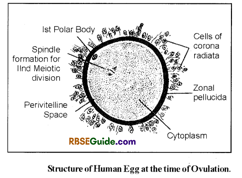
Mammalian Ovum
Mammalian eggs are alecithal having diameter from 100 to 150 micron. It is released at secondary oocyte stage.
Anoncellular. and transparent membrane Zona pellucida is found around cell membrane of egg. At the same time the surface of the oocyte is drawn out into finger like numerous microvilli from the follicle cells in mammals, and this covering of follicle cells is known as Corona radiata .