Rajasthan Board RBSE Class 12 Biology Notes Chapter 2 Male and Female Gametophyte: Structure and Development
In angiosperm plants, flower is formed as a reproductive structure. Morphologically flower is a modified shoot in which the tip of pedicel and nodes and intemodes are highly condensed. Sterile and fertile floral leaves are arranged at the nodes. Hence the flower is also called sporophyll bearing shoot. Normally a typical flower has four types of appandages which are arranged in whorls. These whorls are- Calyx, Corolla, Androecium and Gynoecium. The individual unit of calyx are sepals, of corolla petals, of androecium stamens or microsporophylls, and of gynoecium carpels or megasporophylls.
Structure and Development of Male Gametophyte
Stamen and Microsporangium
In angiosperm plant, male reproductive part is stamen which is known as Microsporophvll. Each stamen is dividec into three parts
- Filament
- Connective
- Anther
Filament is long and slender part, of the stamen, on the top of which it contains anther. Each anther is usually made up of two lobes (anther lobes) connected by a connective. In turn each anther lobe contains two pollen chambers or pollen sacs placed longitudinally. Each pollen chamber represents a microsporangium and is filled with a large number of pollen grains or microspores.
![]()
Thus atypical anther consists of four microsporangia or pollen chambers. This type of anther is known as dithecous. In Malvaceae monothecous condition is found i.e. anthers bear only one lobe (theca with 2 microsporangia) and only two microsporangia or pollen chambers. The microspores or pollen grains develop in the microsporangia
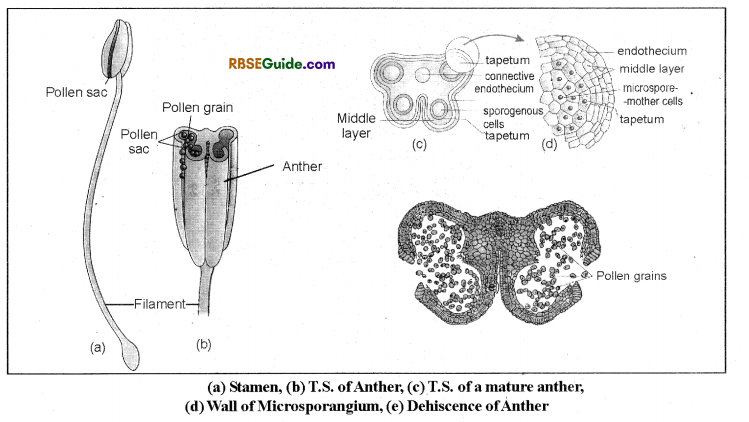
In the early stage of development, the outermost layer of wall of micro sporangium is epidermis and it covers undifferentiated group of homogeneous cells. After structural changes formation of four chambers takes place. The development of microsporangia is Eusporangiate type. Certain cells of hypodermal layer in each lobe begins to enlarge and protoplasm becomes denser than the neighbouring cells. These are archesporial cells or archesporial initials. These cells divide periclinally to form
(i) Primary parietal cells towards outside
(ii) Primary microsporogenous cells towards inner side.
The cells of primary parietal layer usually undergo further periclinal and anticlinal divisions to form a series of 3 to 5 concentric layers forming the wall of the anther. The primary sporogenous cells separate from one another and either develope directly as microspore mother cells or divide mitotically to form sporogenous cells. The last crop of sporogenous cells function as microspore mother cells. So each pollen sac divides into two distinct zones after development and differentiation as
- Anther wall
- Sporogenous cells
Anther wall
Anther wall is differentiated into four layers at maturity stage. These four layers are arranged in a specific order from outer layer to centre, which are as follows
(i) Epidermis
(ii) Endothecium
(iii) Middle layers
(iv) Tapetum.
(a) Epidermis : The epidermal cells of the wall of anther become stretched and flattened as the anther enlarges. It provides protection to anther.
(b) Endothecium : The cells of parietal layer lying below the epidermis are thick walled and radially elongated. Internal wall has deposition of a-cellulose and form fibrous bands which are U-shaped. These cells are hygroscopic in nature and in dry condition stress or tension is created which leads to dehiscence of anther.
(c) Middle Layers: Beneath the endothecium are present two to three layers of cells called middle layers. These cells are thin walled.
![]()
(d) Tapetum: The innermost wall layer, situated just inside the middle layers is.called tapetum. The tapetum surrounds the sporogenous tissue. The cells ofthis layer divide freely and form a layer of enlarged cubical cells around the sporogenous tissue. As the microsporogenous tissue develops, the cells of tapetum elongate centripetally, become multinucleate and get filled with dense cytoplasm.
On the basis of nature of cells, the tapetum can be amoeboid or plasmodial and secretoy or glandular type Most dicotyledenous plants have secretory or glandular type of tapetum.
The rounded structures of fatty nature found in the tapetal cells, are called Probisch bodies. These are converted in to Ubisch bodies by deposition of sporopollenin.
Sporopollenin is most resistant polysaccharide and is an important constituent of the outer wall layer (Exine or exospore) of pollen grains. As the process of microsporogenesis progresses, the cells of the tapetum and the middle wall layers dissolve and contribute to the nutrition and auxin requirement of dividing microspore mother cells. It has been observed that if the cells of tapetum degenerate prior to the formation of microspores or pollen, then the pollen grains fail to attain maturity and may become sterile or abortive.
Sporogenous cells
Mass of cells enclosed by anther wall in pollen sac is called sporogenous cells. These cells form microspor’e mother cells. Each microspore mother cell divides meiotically to form four microspores.
Microsporogenesis
Generally all the microsporogenous cells are capable of producing the microspores but some of them disintegrate and are absorbed by remaining cells. Microspore mother cells divide to form microspores by reduction divisions and the process is known as microsporogenesis. Each activated microspore mother cell divides meiotically to produce four microspores or pollen grains which are arranged in the form of a tetrad.
The cytokinesis may be of successive type or simultaneous type. In successive cytokinesis each of the two nuclear divisions are followed by the formation of wall. In this type the cell plate is formed in the center and then extends centrifugally on both sides dividing the cell into four equal parts. In simultaneous type, division of cytoplasm occurs only after two nuclear divisions dividing the cell into four parts with the formation of furrows. Generally successive type occurs in monocotyledons and simultaneous type in dicotyledons.
![]()
Generally Pollen tetrads are of five types :
(i) Tetrahedral: Only three microspores are observed when we view from one side and fourth microspores is at the back side. Example: Most dicotyledonous plants.

(ii) Isobilateral : In this all four microspores are arranged in one plane of tetrad. Example: Monocot plants.
(iii) Decussate : In this two + two microspores are arranged perpendicular in such a way that upper two microspores are visible and only one from lower tier is visible. Example :Magnolia.
(iv) T-shaped : In this microspores are arranged in a tetrad in such a way that two microspores are arranged in transverse and two in longitudional plane. Example : Aristolochia.
(v) Linear : All microspores are arranged in single linear fashion. Example: Ilalophylla.
The wall between the microspores is made up of callose. This wall is degraded by enzyme callase as a result of which the microspores separate from each other and become spherical. At this stage these are seen freely distributed in the pollen chamber and are called pollen grains.
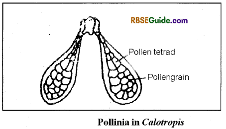
In some plants, pollen grains or microspores of a sporangium cohere (stick) in a single mass called poUiniuin. Example – Calotropis and in some orchids. Most commonly
the microspores soon separate from one another but sometimes they adhere in tetrads to from compound pollen grains, e.g. Drimys, Anona, Drosera, Elodea, Typha. If there are more than four microspores in tetrad then this stage is known as Polyspory.
Dehiscence of Anther:
Breaking of walls of mature anther is called anther dehiscence. The cells of the endothecium opposite the partition between two microsporangia are thin walled and constitute the stomium through which pollen grains are discharged from the pollen chamber.
Anther dehiscence takes place by following methods:
- Transverse dehiscence : When dehiscence occurs transversally. for example Ocimum sanctum.
- Longitudional dehiscence: Dehiscence of anthers occurs longitudionally e.g. Datura, Gossypium.
- Poricidal dehiscence : Dehiscence through one or more small holes (pore) is referred to as poricidal dehiscence. Example : Solanum nigrum.
- Valvular dehiscence : It involves a horizontal opening that causes a lid to separate completely.
Example Berberis.
Structure of Pollen grain :
Each pollen grain is unicellular uninucleated, non dividing and normally round in shape. Its nucleus and dense cytoplasm is covered by two layered wall. Outer layer is called exine and inner layer is called intine. Exine is thick rough and has many different types of ornamentation. These ornamentations may be mesh type, striped or thorny. The exine is rich in a compound known as sporopollenin. which is waterproof, resistant to deterioration and very stiff. This shell is basically one of nature’s most advanced polymers. It ensures that the tender cells inside have a strong chance of survival.
![]()
There are small pore like structures present on exine known as germ pores. Generally the number of germ pores may be one to three. Pollen grains germinate from this germ-pore. Intine is thin, soft and folded structures, which is basically made up of cellulose and pectin. The pollen grains of insect pollinated plants have deposition of oily layer on their surface. This is known as pollen kit. This imparts a specific colour, odour and stickyness to these pollen grains. The substances required for formation of pollen kit are secreted by tapetal cells.
Although function of pollenkit is not clearly understood, scientist believe that it has some role in attracting insects and their sticky nature helps in attaching the pollen with the body parts of the insects. The pollen kit may also protect pollen from ultra-violet rays. Some proteins deposited by tapetal cells and also formed by the pollen are found in the exine of pollens. These are released as soon as pollen grains come in contact with moisture. These proteins are responsible for causing pollen allergy in humans.
Male Gametophyte
Germinated pollen grain is called male gametophyte. Microspore or pollen grain is the first cell of male gametophytic generation. Microspores or pollen grains through the process of gametogenesis form male gametophyte. In this process nucleus of pollen grain divides to form a small lenticular generative cell and a bigger vegetative cell. The generative cell forms two male gametes by mitotic division.
Development of Male Gametophyte:
Microspore is the first cell of male gametophyte. This cell is filled with dense cytoplasm and a distinct nucleus. After being separated from tetrad it increase in size. Cytopolasm appears as single layered villi like and many vacuoles are formed. Some of the stages of development of male gametophyte are completed before pollination and some after pollination. Before pollination, nucleus of pollen grain divides by mitosis into large vegetative nucleus and small generative nucleus and then by formation of wall between these two nuclei leads to formation of larger vegetative cell and a smaller generative cell.
![]()
Generally in most of the plants pollination occurs at this two nucleated stage. The pollen grain after liberation from pollen sac reach the stigma of flower. Intially generative cell is attached with wall of pollen grain, but after formation of complete wall it separates from wall of pollen grain and moves towards vegetative cell. After separation from pollen wall, generative cell becomes flattened and later on may become elliptical, lenticular or even spindle shaped.
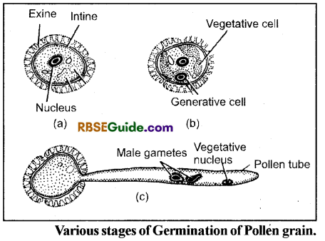
Vegetative cell:
Size of vegetative cells is large. Its nucleus is initially round shaped and later on becomes undefined shaped. There is increase in shape and size of various organelles. It contains adequate amount of RNA and proteins as the vacuoles disappear. Pollen tube is formed by vegetative cell probably at the time of germination.
Generative cell: Initially generative cell is lens shaped and after separation from wall of pollen grain it becomes round shaped. At initial stages of development it’s shape changes. Generally it appears elongated like worm shaped. Their longer shape allows their easy movement in pollen tube. Generative cell contains all organelles and cytoplasm is found in very small quantity.
Both, generative and vegetative cells have equal amount of DNA but amount of RNA is different in cytoplasm. Amount of RNA is more in vegetative cell as compared to generative cell because of large amount of cytoplasm in vegetative cell.
Formation of Male gametes :
Male gametes are formed by division of generative cell. In some plants mitotic division of generative cell takes place after pollination but prior to germination of pollen grain. Example: Portulaca, Haloptelea etc. However in most angiosperms the generative cell divides by mitotic division in the pollen tube just after the germination of the pollen grains. The pollen tube grows through the stigma and the vegetative nucleus moves in the pollen tube and after this the generative cell moves in the pollen tube.
Here by the division of generative cell, two male gametes are formed. Thus in majority of angiosperms male gametes are formed in the pollen tube while it is growing through the stylar rergion. Example: Lilium, Nimophilla etc. Each male gamete is unicellular, non motile, and uninucleated structure.
Note: In majority of angiosperms, pollination takes place at two celled stage of pollen grains and two male gametes are formed in the pollen tube while it is growing through the stylar region
Structure and development of Female Gametophyte
Carpel or Megasporophyll: Female whorl of flower of angiosperm plant is known as Gynoecium. Gynoecium is formed from one or more carpels. Carpel is a modified leaf which bears ovules and is called as megasporophyll. When the number of carpels is more than one and are present individually, then condition is called apocarpous and when they are fused together the condition is caled as syncarporus.
Carpel is flask shaped structure and it can be divided into three parts as :
- Stigma,
- Style and
- Ovary.
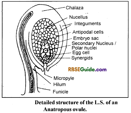
Ovary is swollen, basal, hollow part of gynoecium which contains many small spherical structures, called as ovule. Ovule is also called megasporangium. Each ovule is attached to the inner wall of ovary on placenta. This attachment to the placenta is by slender stalk known as funicle.
![]()
The point of attachment of the body of the ovule to its stalk (funicle) is known as hilum Sometimes the funicle continues beyond the hilum, along the body of the ovule forming a sort of ridge which is called raphae.
Elongated slender part of ovary is called style. This provides pathway to pollen tube. Stigma is present on top most part of style and it receives pollen grains at the time of pollination.
Structure of Ovule:
Structure of ovule is complex in angiosperm plants. The mature ovule is almost round in shape and differentiated into the following parts. The main body of ovule is made of parenchymatous tissue called as nucellus (2n). Enclosing the nucellus are 1 or 2 integuments (2n). Outer-one is called as outer integument and inner one as inner integument. If there are two integuments in the ovule it is called bitegmic, if there is only one integument the ovules is called Unitegmic (Sunflower, Tcigetus).
Some ovules do not have any integuments and are called ategmic (Loranthus, Santalum). The integuments are incomplete at the apex of ovule forming a pore called micropyle. The base of the ovule from which integuments arise is called chalaza. Acentrally located large cell (female gametophyte) with eight nuclei is called embryosac. Three of the nuclei present at micropylar end of the embryo sac are called egg apparatus, which consists of one egg cell (central cell) and two laterally placed cells called synergids.
At the chalazal end of the developing embryosac, there are three antipodal cells. In the centre of embryo sac are present two, polar nuclei, which just before fertilization fuse to form
Types of ovule:
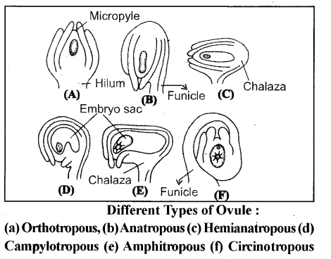
1. Orthotropous ovule: “The micropyle, chalaza and funicle lie in one straight line. E.g., family polygonaceae, piperaceae and ovule of most gymnosperms.
2. Anatropous (Inverted) ovule : The body of the ovule is completely inverted so that micropyle and hilum come to lie very close to each other. 82% families of angiosperms contain this type of ovule. E.g. plants of gamopetalae.
3. Campylotropus: When the ovule is curved, the micropyle is directed towards chalaza. Chalaza is situated at right angles to funicle. E.g. Members of cruciferae and leguminoceae family.
4. Amphitropus or Transverse Ovule: The ovule curvature is more pronounced and embryosac becomes horse-shoe shaped eg. Families A hsmaceae, Butumaceae.
![]()
5. Hemianatropous Ovule : When nucellus and integuments lie more or less at right angles to the fimicule. E.g. FamiliesRanunculaceae and Primulaceae.
6. Circinotropous ovule : The funicle is exceptionally long and forms a complete circle around the ovules, which is free from it except for a small area at the end of funicle. E.g. Opuntia and other members of families Cactaceae and Plumbaginacea.
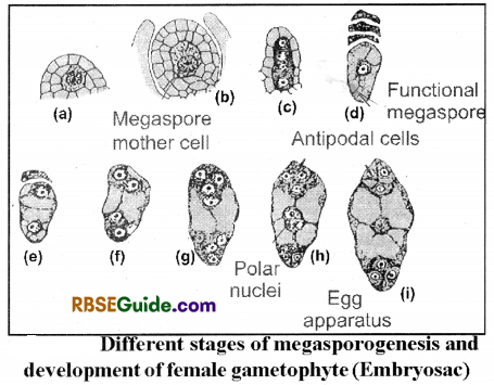
Megasporogenesis
Archesporium : Either one hypodermal cell at the apex of nucellus functions directly as megaspore mother cell or the hypodermal cell first divides unequally into outer parietal and an inner primary sporogenous cell. The parietal cell in this case may form a variable number of wall cells. The primary sporogenous cell is large, has a distinct nucleus with dense cytoplasm. Primary sporogeneous cell does not divide and acts as megaspore mother cell.
Primary outer parietal cells either does not divide and if divides it may devide many number of times to form wall. The first meiotic division of megaspore mother cell is always transverse forming a dyad of cells. Second meiotic division is also transverse resulting in a linear tetrad of four megaspore. Linear tetrad of megaspores is most common four but T-shaped, 1 -shaped or isobilateral tetrads may also form.
Generally the lowermost megaspore (farthest from micropylar end) is functional i.e., develops into embryosac or female gametophyte. Other three megaspores degenerate and being consumed for nutrition by functional megaspore. Development of Female Gametophyte or Embryo sac
The first division of functional megaspore gives rise to two nuclei, the primary micropylar and primary chalazal nuclei. The second division results in one pair of nuclei at each end, and the third division produces four nuclei at one end and four on the other end (i.e., at opposite poles of the now elongated developing embryo sac). Three of the nuclei at the micropylar end become organized to form egg apparatus and one nucleus which does not participate in the formation of egg apparatus and is called polar nucleus.
![]()
At the opposite (chalazal) end of the developing embryo sac, three nuclei differentiate as antipodal cells (or nucleus) and the fourth nucleus fonns the polar nucleus. The two polar nuclei from opposite ends (mieropylar, and chalazal) migrate in the centre of embryo sac giving rise to secondary nucleus ( = definitive nucleus). Most ofthe angiosperms have this type of embryo sac development and is known as normal Type or Polygonum type. When the embroy sac develops from one megaspore, it is called as monosporic embryo sac. Structure ofthe Mature embryo sac
A typical mature embryo sac consists of a large cell with two polar nuclei (which on fusion give rise to secondary nucleus in the centre). At the mieropylar end there is an egg apparatus (1 egg cell with 2 synergids) and at its chalazal end are found 3 antipodals. The cells of egg apparatus and antipodal cells are uninucleate and haploid but the central cell is first binucleate. The two polar nuclei later on fuse to form a diploid nucleus called secondary nucleus = definitive nucleus).