Rajasthan Board RBSE Class 12 Biology Notes Chapter 30 Man-Movement & Locomotion
General
Skeleton forms the basic supporting framework of the body. The skeleton originates from embryonic ectoderm & mesoderm.
In the vertebrates, the skeleton is of two kinds
Exoskeleton
- It is found outside the body’. It derives from epidermis or
- dermis or from both layers. Normally the exoskeleton is
- dead but it may be living.
- Example-scales in fishes, scales and scutes in reptiles,
- feathers & claws in birds and horns nails and hairs m mammals.
![]()
Endoskeleton
Endoskeleton is located inside the body. It is composed of bones and cartilages. It derives from embryonic mesoderm.
It is living.
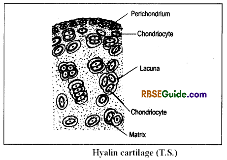
Cartilages and Bones
- The cartilages are soft & elastic.
- It contains matrix & chondrioblasts cells.
- The matrix is made up of chondrin, protein & collagen fibres. In some cartilages, the matrix also has elastin fibres & salts.
- The chodrin is a chodro-mucoprotein which provide strength & elasticity” to the cartilage.
- The chondrioblasts form chondriocvtes & the matrix.
- The chondriocvtes remain distributed in the matrix and are enclosed in lacunae.
- Each lacuna encloses one to four chondriocvtes.
- The membrane of the lacuna has blood capillaries which supply O, & food to the chondriocvtes.
- The cartilage is bounded by a perichondrium.
- The cartilages are of 4 types –
(i) Hyaline cartilage
- The matrix is clear, semi-transparent & light blue in colour.
- It is elastic.
- The matrix is without fibres.
- It is found in hyoid, tracheal rings, larynx and ends of bones of legs & ribs.
(ii) Elastic cartilage
- It’s matrix has yellow elastin fibres which are branched.
- It is found in pinna, epiglottis, & tip of nose.
(iii) Fibrous cartilage
- Its matrix has bundles of white collagen fibres. They are unbranched.
- It is tough cartilage.
- It forms intervertebral discs & pubic symphoisis of pelvic girdle
(iv) Calcified cartilage
- It is a type of hyaline cartilage in which matrix has CaCO3. Hence, it becomes hard like a bone.
- It is found in pubis of pelvic girdle & supra-scapula of frog.
![]()
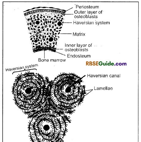
Bones
To study”, the bones are decalcified in HCl because decalcified bones become soft & elastic.
The cavity in the hollow.bone is called as bone marrow. The bone marrow at the ends is called as red bone marrow & in the centre as yellow bone marrow.
The yellow bone marrow has adipose tissue where as in the red bone marrow RBC are formed.
The outer layer the of the bone is called as periosteum and the inner layer as endosteum. The bone has two layers of the osteoblasts viz-inner & outer layers. These osteoblasts form osteocytes & matrix.
The matrix is formed first & it is made up of ossein protein. Lateron there is deposition of calcium salts in the matrix which is called, as calcification.
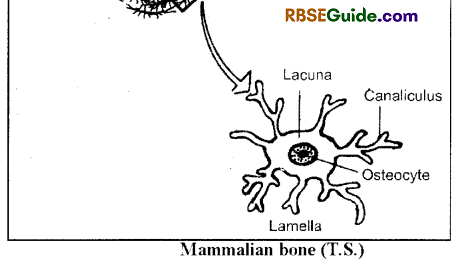
The osteocytes form special Haversian systems or osteon in the matrix.
Each Haversian system has a central Haversian canal that is surrounded by 4 to 20 concentric rings of lamellae.
Each lamella has an osteocvte surrounded by lacuna. The lacuna gives out mam’ minute canaliculi. .All the lacunae of one Haversian system remain interconnected with the help of canaliculi.
The Haversian canal has blood vessels & nerves. The nutrient from the blood reach to all the osteocytes through these canaliculi.
All the Haversian canals remain interconnected & remain connected to the bone marrow with the help of Volkmann’s canals.
Formation of bones is called as osteogenesis or ossification.
The bones contain mainly Ca3(P04)2 and little CaC03 / Mg3(P04)2 NaCl. They make the bone strong & hard.
These mineral form 67% part of the bone.
The bones remain unaffected in the solution of KOH
The bones are of 3 types-
(i) Membranous or Investing bones
- They are formed by ossification in the membranes situated below the embryonic skin.
- They invest soft cartilagenous parts of the skeleton & prov ide them strength.
- Example – Flat bones of skull, clavicle, bones of digits etc.
(ii) Cartilagenous or Replacing bones
- It is formed in place of hyalin cartilage.
- The matrix of the cartilage is destroyed by special osteoclasts.
- Exam pie – Bones of limbs, vertebral column, some bones of skull.
(iii) Sesamoid bones
- It is formed by the ossification in the ligament.
- Example – Patella bone.
![]()
Functions of Skeleton
- The Skeleton forms a rigid frame work and supports the body.
- Skeleton provides a base for attachment of muscles.
- Skeleton acts a lever during various movements of the body and also help in locomotion.
- It protects number of delicate organs such as brain, heart, lungs and spinal cord.
- Blood corpuscles are produced in the red bone marrow
Endoskeleton
Endoskeleton in man is well developed and it is divisible into 2 parts-
1. Axial Skeleton
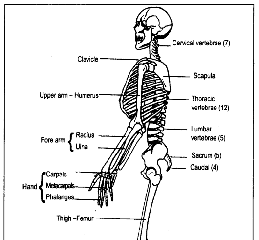
The axial skeleton includes skull, vertebral column, sternum & ribs.
There are 80 bones in the axial skeleton of man.
2. Appendicular Skeleton
The appendicular skeleton includes girdles and limb bones.
There are total 126 bones in the appendicular skeleton of man.
The human endoskeleton in all includes 80 126 206 bones.
Axial Skeleton
Skull
The skeleton of head is called as Skull. The human skull is dicondyllic & consists of 29 bones which are joined together by sutures.
The skull is divisible into 2 parts
(A) Cranial region
(B) Facial region
(A) Cranial Segment
It is the large & back part of the skull which is divisible into 5 parts:
(i) Occipital segment
- It is the basal part of the cranium
- It has foraman of Magnum through which spi¬nal cord comes out.
- The foraman is surrounded by 4 bones viz.
- (a) Basi-occipital One & below the pore
- (b) Supra-occipital One & above the pore
- (c) Ex-occipital Two & both the sides of pore.
- Each ex-occipital has an elevation called occipital condyle. Hence, the skull is dicondyllic.
(ii) Parietal region
- This part has 5 bones
- (a) Parietal bones – 2 & in upper part
- (b) Alisphenoid – 2 & in lateral sides
- (c) Basisphenoid – 1 & in lower part
- Both the parietal bones remian separated by interparietal bone.
- The basisphenoid has a depression called sella tursica that lodges the pituitary gland.
(iii) Frontal region
- It has 5 bones viz.
- (a) Frontal – 2 & upper
- (b) Pre-sphenoid – 1 & lower
- (c) Orbitosphenoid – 2 & lateral
- An ethamoid bone is attached in front of it which is highly porous
- The frontal bones form supraorbital ridge above the eye orbits.
(iv) Auditory capsule
- This part is situated between the occipital & parietal parts.
- It has 2 bones
- (a) Periotic bone
- It is compound bone which is formed by the fusion of preotic, epiotic & opishotic bones.
- It has 2 parts
- Petrous part – Inner & forms circle around the internal ear.
- Mastoid part – Posterior part & it is porous
![]()
(b) tympanic bulla
It is a flask-shaped bone. It’s mouth is attached with pinna. It’s neck forms external auditory meatus. It’s main cavity is called as middle ear or tympanic cavity.
The middle ear has three ear ossicles
viz. malleus, incus & stapes
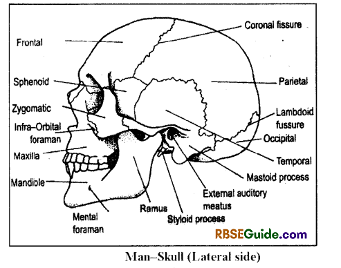
(v) Orbits
It is situated in the frontal region of the skull
It has 2 eye orbits which remain separated by an inter-orbital septum.
The skull with interorbital septum is called as tropibasic
There is a small lacrymal bone in front of eye orbit which has a groove for lacrymal duct.
(B) Facial part
This part includes olfactory capsule, upper j aw & lower jaw.
(i) Olfactory capsule
This part has 7 bones:
(a) Vomer – One, long, thin and lower bone.
(b) Nasal – Two, flat and upper bones
(c) Pre-maxilla – Two, both sides
(d) Maxilla – Two, both sides
It has two nasal chambers which remain separated by internasal septum or mesethmoid.
Each nasal chamber has a turbinal bone which is porous & scrolled.
The turbinal bone has 2 parts
- (a) Ethmoturbinal – attached to ethmoid bone.
- (b) Nasoturbinal – attached to nasal bone
(ii) Upper jaw
It has 2 equal halves and each half includes 6 bones –
(a) Pre-maxilla:
Anteriormost bone which has two incisor teeth.
It forms snout in rabbit
(b) Maxilla:
It is attached with three premolars & three molars both are together called
as cheek teeth.
It’s posterior part has a palatine process
(c) Palatine:
It is situated just behind the palatine process.
(d) Pterygoid:
It is a small bone which is attached to the palatine
(e) Jugal:
It is a narrow & wavy bone
It forms zygomatic arch by joining the processes of maxilla & squamosal
(iii) Lower jaw
- It is also called as mandible.
- It has two equal parts which remain united by a symphoisis.
- Each part has a dentary bone
- Each dejitary has one incisor, two premolars & three molars.
- In rabbit, the gap between the incisor & premolar is called as diastema.
- The back part of the dentary forms a condyle. There is coronoid process in front of condyle & an angular process behind the condyle
- The lower jaw is attached to the squamosal bone. The attachement of the lower j aw is called as cranio-stylic suspension.
![]()
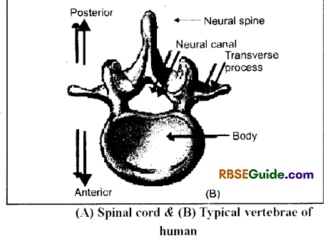
Vertebral Column
Vertebral column is a salient feature of all vertebrates. It is found mid-dorsally from head to tail.
The vertebral column is made of vertebrae. The salient
features of mammalian vertebrae are as follows-
- The centrum of the mammalian vertebrae is acoelous.
- The centrum has a bony plate on both the anterior & posterior parts which is called as epiphysis.
- There is an intervertebral disc between two vertebrae which is made up of fibrous cartilage.
The vertebral column has 5 parts –
- Cervical region
- Thoracic region
- Lumbar region
- Sacral region
- Caudal or Coccygeal region
There are 33 vertebrae in man & the vertebrae formula is c7, T12, L5, S5, Cd4
(A) Cervical vertebrae
There are 7 cervical vertebrae.
First cervical vertebra is called as atlas, second as axis and 3rd to 7th are called as typical cervical vertebrae.
(i) Atlas:
It is a ring-like vertebra which holds the skull
It is without centrum & zvgapophysis. The transverse processes are long, flat & wing-like.
Neural spine is very small and neural canal is large.
(ii) Axis:
Presence of odontoid process anterior to the centrum. It forms an axis for head rotation
The transverse processes are small
Neural spine is flat & ridge-like.
Absence of pre-zygapophysis
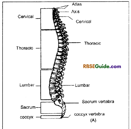
(iii) Typical cervical vertebrae:
3rd to 7th cervical vertebrae are called as typical vertebrae.
The centrum is broad.
Neural spine & neural canals are developed. The neural spine of 7th vertebra is more longer.
Each vertebra has two vertebraterial canals
Each is fused with a bifid rib
Presence of zygapophysis.
(B) Thoracic vertebrae
- There are 12 thoracic vertebrae
- Their centrum is well developed.
- Neural spine in first 9 vertebrae is long, thin & posteriorly directed. In rest, the neural spine is small & straight.
- They are attached with ribs,
- The ribs of first 9 vertebrae are long & large and of last three vertebrae are small & floating.
![]()
(C) Lumber vertebrae
- They are comparatively strong, large & heavy.
- The centrum is long
- Presence of developed neural canal, neural spine & zygapophysis.
- They have stout transverse processes which are downwardly pointed
- In addition, they have anapophvsis & metapophysis
- First two lumbar vertebrae have hypophysis on the lower side of the centrum
(D) Sacral vertebrae
- Rabbit has 4 sacral & man has 5 sacral vertebrae. They fuse to form a sacrum.
- The sacrum is situated in the middle of the pelvic girdle & it remain attached to the ileum.
- First sacral vertebra is stoutest. It’s centrum is thick & flat. It’s transverse process & sacral ribs fuse to form 4 fiat wing like processes.
- The neural spines remain posteriorly directed.
(E) Caudal or Coccygeal vertebrae:
There are four very small caudal vertebrae
Sternum or Breast Bone
It forms respiratory basket along with ribs & thoracic vertebrae
It is a flat bone which has distinct flat anterior & posterior sides.
It has 3 parts
- Manubrium – Upper part; the clavicle & ribs are attached to it.
- Sternum body or Glandiolous – Middle part, 2nd, 3rd, 4th 5th, 6th & 7th ribs get attached to it.
- Xiphoid process or Ensiform – Posterior part, small & triangular plate.
The ribs articulate its ends.
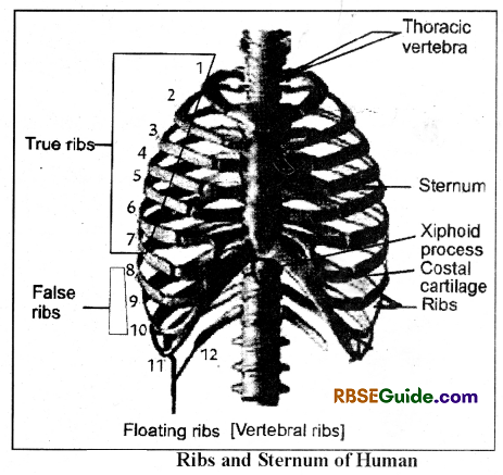
Ribs
The man has 12 pairs of ribs. They are of 3 types:
(i) True ribs-First seven pairs of ribs are called as true ribs. They are attached separately to the sternum.
(ii) Flase ribs – 3 pairs (8th, 9 & 10th) are called as false ribs. They are not attached to the sternum directly but are attached to the sternal part of the 7th rib.
(iii) Floating ribs – 2 pairs (11th & 12th pairs) are called as floating ribs. They are small & never reach to the sternum.
Each rib is bicephalous i.e. it has two heads:
(a) Tuberculum-It is attached to the transverse process of the thoracic vertebra
(b) Capitulum It is attached to the centrum of the thoracic vertebra
The tuberculum of floating ribs are small and are not attached to the vertebra
Appendicular Skeleton
It is situated on both the sides of the longitudinal axis of the body.
It includes girdles & bones of fore linbs and hind linbs.
The girdles are of two types-Pectoral girdle & pelvic girdle.
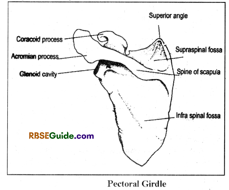
Pectoral Girdle
- It supports fore limbs & protects chest organs.
- It has two equal halves.
- Each half has a clavicle & a scapula-coracoid bones
- The clavicle is a rod-like bone which is also called as collar bone.
- The scapula is a broad & flat bone which is found in all mammals.
- The coracoid is get fused with the scapula & found in the form of a coracoid process.
- The supra scapula is absent in the pectoral girdle of Man.
- The dorsal side of the scapula has a spine. The anterior & ventral extension of the spine is called as acromian process
- The posterior process of acromian in the rabbit is called as metachromian process.
- The head of the scapula-coracoid has a cavity, the glenoid cavity. The head of the humerus bone articulates to the glenoid cavity.
![]()
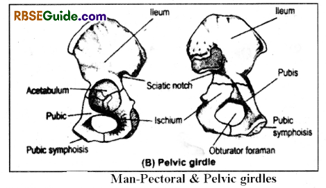
Pelvic Girdle:
It supports hind limbs.
It has 2 equal halves & each half is called as osinnominatum.
Both the os-innominata remain united by pubic svmphoisis which is made up of fibrous cartilage.
The pelvic girdle of rabbit is W-shaped.
Each os-innominatum of rabbit has 4 bones – Ilium, Ischium, Pubic & Cotyloid
Each os-innominatum has a groove, the acetabulum. The head of femur articulates with it.
In rabbit, the pubic bone does not participate in the formation of acetabulum. There is a small cotyloid bone between the acetabulum & the pubic.
In man, cotyloid bone is absent. Hence, each os- innominatum has 3 bones-Ileum, Ischium & Pubic. All of them participate in the formation of acetabulum.
There is an obturator foraman between pubic & ischium bones.
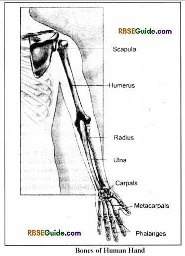
Fore limb has 5 parts:
(A) Upper arm
(B) Fore arm
(C) Wrist
(D) Palm
(E) Fingers
(A) Upper arm
IPs bone is called as humerus.
It is a cylendrical & strong bone which has distinct proximal & distal ends.
The proximal end is called as head which articulates to the glenoid cavity. It also has a greater tuberosity & a lesser tuberosity on the lateral sides.
It’s distal end has a pullv-like trochlea, an olecranon fossa & a sura- trochlear foraman.
It has a deltoid ridge which is a ridge from proximal end to the middle part on the ventral side.
(B) Forearm
This part has two parts viz- Radius – inner & thick and uina – outer & comparatively long.
At the proximal end, the ulna extends to form olecranon process & a sigmoid notch. These structures form elbow joint with the trochear part of humerous. At the distal end. it has two elevations called epiphysis.
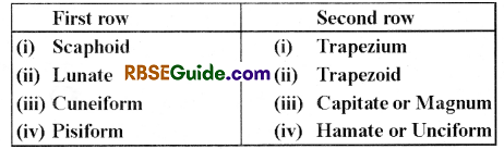
(C) Wrist
The wrist of man has 8 carpals which are arranged in two rows 4 and 4.
(D) Palm
Palm of both rabbit and man has 5 – 5 long bones called metacarpals.
(E)Fingers
The fore limb of both rabbit & man has 5 – 5 fingers. Their bones are called as phalanges.
First finger is called as thumb which has 2 phalanges. Remaining 4 fingers has 3 – 3 phalanges.
The tip of each finger has a claw in rabbit & nail in man.
The claws a& nails are made up of keratin.
![]()
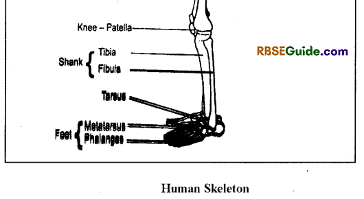
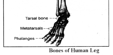
Bones of Hind leg
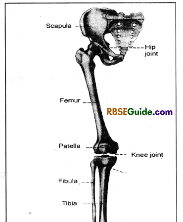
The hind leg has 5 parts viz
(A) Thigh
(B) Shank
(C) Ankle
(D) Foot
(E) Toes
(A) Thigh
- This part has a femur bone which is the longest & stoutest in the body.
- It has 2 distinct ends and a middle shaft.
- It’s proximal end has a head, a greater trochanter, a lesser trochanter & a third trochanter»
- The head articulates with the acetabulum. It’s distal end has a patellar groove intercondyllar groove.
(B) Shank
- This part has 2 bones viz.- tibia & fibula. Their proximal ends fuse to form a tibio-fibula bone.
- The tibia is a long, straight & strong bone.
- The fibula is a small & thin bone.
- The proximal end of tibia has a cnemial crest.
- It’s proximal end forms knee joint with femur. The knee joint is covered by patella bone which is also called as knee cap.
- It is a flat & triangular bone which is formed by the modification of tendon. Hence, it is a sesamoid bone.
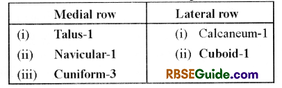
(C) Ankle
- It is formed by tarsals bones.
- Man has 7 tarsals which are arranged in 2 rows of 5 + 2.
(D) Foot
- This part has metatarsal bones
- Foot of rabbit has 5 metatarsals
(E) Toes
- Rabbit lack first toe or hallux. Remaining 4 toes has 3 + 3 + 3 + 3 phalanges.
- Man foot has one hallux & four toes. They have 2 + 3 + 3 + 3 + 3 phalanges.
- The lips of toes have claws in rabhit & nails in man
Joints
- In the vertebrates, joints are the places where two bones articulate with each other or bone and cartilage articulate with each other. In other words the contact place between two or more bones or between bone and cartilage is known as Joint.
- These joints facilitates movements of the body parts.
- On the basis of movements, the Joints are of three types-
Synarthrosis or fixed or Immovable Joints
- The articulating bones are held together by a dense bundle of tough and white fibrous connective tissue which cannot be stretched or extended.
- No movement is possible and hence called as fixed joints or synarthrosis
- Examples-Joints between the bones of skull which are called Sutures.
Amphiarthrosis or partially movable joints
- It is a tough joint in which bones are joined by a disc of white fibrous cartilage which can be stretched a little.
Limited movement is possible - Example – Pubic Symphoisis
- It is of two types-
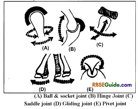
(i) Pivot Joint – Joints between Atlas and axis vertebrae. It provides lateral movement.
(ii) Gliding Joint – In suchjoint, end of one bone glides over a certain portion of another bone. Movement in different directions is found. Joints between tarsal bones of ankle, carpal bones in wrist and in between sternum and clavicle.
![]()
Synovial or Diarthrosis ou movable Joints
The Bones joined by synovial joints can move in one or more directions freely. Highly smooth thin layer of hyaline cartilage is found on the surface of the bone where joint occurs. This cartilage minimize the friction between the bones. A space is found between the joint of bones, which is called synovial cavity. Bones are joined with each other by many ligaments.
Ligaments fuse together and form a capsule which is covered by synovial membrane. It secretes mucin containing synovial fluid. This fluid provides nutrition to hyaline cartilage and lubrication to joint
It is of three types-
(i) Ball and Socket Joint – It this j oint ball like end of one bone fits into the socket of another bone. Movement of bone with the ball head in various planes is possible. Example-Shoulder Joint and Hip Joint.
(ii) Hinge joint – Resemble the hinges of doors. Movement in one direction only. Examples – Elbow joint, knee joint and finger joint, occipital condyle and atlas joint.
(iii) Ellipsoidal joint – Movement of one bone over the another bone, allowing movements in around two axis. Joints between metacarpals and phalanges in hand, and metatarsals and phalanges in foot. Another example is joint between radius and
Muscles
General
All the higher animals needs muscles to perform various movements & locomotion. The muscles also provide a characteristic posture to the body.
The muscles are formed by the epimeric & hypomeric layers of the mesoderm. The epimeric layer finally forms voluntary or striated muscles. The splanchnic layer of the hypomeric layer forms involuntary or unstriated muscles.
Study of muscles is called as myology or sarcology.
Muscles exhibit contractabilitv.
The muscle cell is also called as muscle fibre.
The intercellular space is absent in the muscles.
Types of muscles
The muscles are of 3 types-
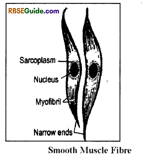
Unstriped muscles
They are also called as involuntary muscles. These muscles are found in the wall of alimentary canal, testes & their ducts, ureters, urinary bladder & blood vessels. This muscle consists of spindle shaped & long cells.
Each cell (muscle fibre) has a large central nucleus. This nucleus is surrounded by cytoplasm which is called as sarcoplasm.
The sarcoplasm has many small threads or myofibrils which are contractile. The myofibrils are surrounded by a thin membrane & they are found in many layers.
They are not under the control of will, hence are called as involuntary muscles.
These muscles are inervated by nerve fibres of sympathetic nervous system.
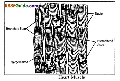
Cardiac muscles
This muscle forms walls of the heart. Their muscle fibres are branched & form a network.
The muscle fibre is bounded by a sarcolemma & has light and dark bands.
In addition, the muscle fibre has transverse plates, the intercalated discs. The intercalated discs provide false appearance of cells in the muscle fibres.
The cardiac muscles are involuntary. They never exhibit muscle fatigue and contract rhythmically.
Striped muscles
These muscles are voluntary and they form the muscular part of the body.
They always remain attached to the skeleton. Hence, they are also called as skeletal muscles.
Their muscle cells are long, cylindrical and bounded by sarcolemma. Each muscle fibre is multinucleated, hence also called as syncytial.
Functional Architecture of skeletal muscle
General
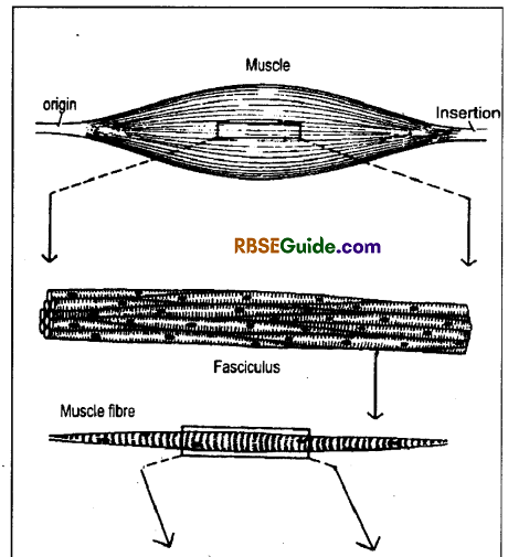
Each skeletal muscle is spindle-shaped & has 2 distinct ends viz.-Origin & Insertion. The origin & insertion remain attached to the bones with the help of tendon (cord-like structure) or aponeurosus (membrane-like structure).
The skeletal muscles of the tongue & upper part of oesophagus are not attached to the skeleton.
These muscles are also called as fatigue muscles or phasic muscles or striated muscles or somatic muscles.
Their structural unit is called as muscle fibre.
The muscle fibre is multinucleated or syntitial & bounded by sarcolemma.
The muscle fibres are found in group & each group is called as fasciculus. The fasciculus is bounded by endomycium.
The fasciculi are also found in the group and each group of fasciculi is bounded by perimycium which is made up of connective tissues.
The whole skeletal muscle is bounded by an epimycium.
Fine structure of muscle Fibre
The maximum length of the muscle fibre is 30 cm (1 – 30 cm) and the diameter is 0.01 to 0.1 mm. It is bounded by a sarcolemma or plasmalemma or myolemma. It’s Sarcoplasm has many mitochondria which are called as sarcosomes. The ER found in the sarcoplasm is called as sarcotubules.
The sarcoplasm has three soluble proteins viz.myogen, myoglobin & myoalbumin.
![]()
The sarcoplasm stores glycogen. In addition, it has less quantity of Na, Ca, P & Mg & more quantity of K.
The muscle fibre has many myofibrils. The length of the myofibril is similar to the muscle fibre but it’s diameter is 1 to 3 μ.
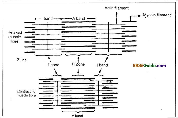
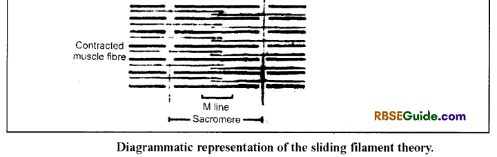
The Myofibril has alternate A-bands (Anisotrophic) & I – bands (Isotropic).
There is a Z – line (zigzag) in the middle of the I. band which is also called as Krau’s membrane.
The middle zone of the A-band is called as H- zone which has a central M-line.
The distance between two Z-lines is called as sarcomere which is a functional unit of the muscle. Each sarcomere includes one complete A – band and 2 halves 1-bands on both the sides (I/2 + A + I/2).
The A-band is made up of mainly myosin filaments. The length of the myosin filament is 1.5 μ & the diameter is
100Å.
The I – band is made up of mainly actin filaments. The length of actin filament is 2μ & the diameter 50Å.
0.2μ of both the ends of the actin filaments siozue remain in the A-bands.
During resting phase, the length of the A-band is 1.5μ & of half I-band is 0.8μ. Hence, the length of the sarcomere is 3.1μ.
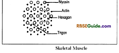
In the transverse section of myofibril, through the end of A-band, has 5000 filaments (Both actin & myosin). Each myosin filament is surrouned by 6 actin filaments (Hexagon). Similarly one actin filament is surrounded by 3 myosin filaments (Trigon)
The myosin filaments are provided with cross links or bridges. The cross links remain directed towards the actin. They are attached at an angle of 60° & are equally spaced by 60 – 70 Å The H- zone is without cross-links.
![]()
Molecular Organization of the contractile System
General
There are three types of proteins in the contraction Process-
1. Force Generating Proteins:
Examples-
(a) Myosin
(b) Actin
2. Regulatory Proteins:
Examples-
(a) Tropomyosin
(b) Troponin
3. Structural Proteins:
Examples-
(a) a Actinin
(b) M-protein
(c) C-protein
Myosin:
Myosin forms primary and thick myofilament. Approximately half part of protein in a filament is of myosin. Its molecular weight is 500,000. It is an a-helix protein.
Actin
Its molecular weight is 42000. Actin is found in two forms- Glactin and Factin. Many units of globular actin – G- actin unite and a chain of filament actin F-actin is formed. In muscle, fibre actin is always occurs in F-actin form, which is a polymer of G-actin. It is found in the form of two coiled strands.
Tropomyosin:
Its molecular weight is 64000. It is made up of two a- helix units, which are approximately 400A long.
Troponin:
The other protein joined with tropomyosin is troponin. It is of three types-i.e. Troponin-C, Troponin – I and Troponin – T.
- Troponin-C-Calcium binds with this protein. Molecular weight is 17000.
- Troponin-I-It acts as inhibiter for the bonding of myosin and F-actin. Molecular weight is 22000-24000.
- Troponin-T-It is linked with tropomyosin. Molecular weight is 24000-37000.
C-Protein
- It is found in the middle of A-band of myosin myofilament. 30.2.4.7 M-Iine protein
- It forms M-line. The role of this protein is to keep the myosin myofilaments in their positions.
Sarcotubular System
It is found in sarcoplasm and made up of following structures:
(i) T-system-This system contains transverse tubules. They have direct connectivity with Sarcolemma.
(ii) Sarcoplasmic reticulum-Around each myofilament, shiny endoplasmic reticulum modified into Sarcoplasmic reticulum. It is mainly made of minute tubules and cistemae.
Sarcosome – Mitochondria of muscle cells are called Sarcosome. Size of Sarcosome is bigger in comparison to the mitochondria of other cells.
Neuromuscular Junction
It is a physiological communication between muscle & nerve fibre. The nerve cell axon which innervate the skeletal muscle is called as motor neurone. This axon is myelinaed and divides near the muscle fibre to form thin branches which are non myelinated. These branches are very closely sitated near the sarcolemma.
Their terminal ends form knoblike structures called synaptic knobs. They are full of acetylcholin (Ach) as neurotransmitter. The muscle cell modified to form motor end plate which bear Ach receptors.
Mechanism of Muscle Contraction
Sliding Filament Theory
- The process of muscle contraction can be explained with the help of “Sliding filament theory” which was given by A.F. Huxley, J. Hensen & H.E. Huxley.
- According to it, the myosin filaments remain stationary & the actin filaments slide over the myosin with the help of cross links.
- During contraction, the length of the A-band remains unchanged but the length of the I-band & H-zone get reduced.
- In H-zone, the actin filaments slide over each other. The contraction results in the shortning of the
- The contraction results in the shortning of the sarcomere.
- The relaxation of the muscle involves reverse sliding of the actin filaments to obtain original position.
- The process of muscle contraction & Relaxation involves four phases:
![]()
(A) Excitation:
Nerve impulse promotes releasing of Neurotransmillar Acetylcholine the ends of axon at Neuromuscular junction. This chemical changes permeability in plasma membrane of muscle towards Na+.
Due to this, Na+ enters into muscle cell and causes change in charge (axon potential), which is +ve inisde plasma membrane. Innormal condution, there is -Ve charge inside the inner surface of plasma membrane. This positive change is transferred on whole plasma membrane of muscle and generates action potential, which is known as Excitation state of muscles.
(B) Excitation – Contraction Coupling :
This is the process in which action potential promotes contraction in muscle cell. This action potential instantly transfers to T-system,
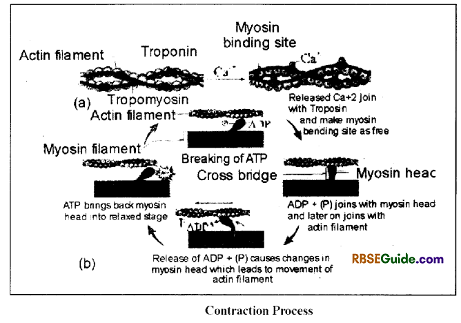
Due to this potential, Ca++ released from sarcoplasmic reticulum and join with troponin C. It results in configurational changes in Troponin atoms. As a result, tropomyosin and troponin, both are released from active site of actin. As soon as active site is made free the myosin filament joins with actin and contraction process starts.
(C) Contraction
Contraction occurs by sliding filament process, which is initiated by joining of actin & myosin transvers bridge.
Just before joining of active filament at active site, one atom of ATP joins’ at head of transverse bridge. ATP present in myosin head breaks down into ADP+ Pi by ATPase.
ADP+ Pi remain attached or joined with head after that myosin head joins with the active site of the actin filament.
There are conformational changes in the head due to joining of head and active site. Due to this, there is bending in head (just as we bend our finger) and actin filament is pulled towards the centre of the sarcomere (H zone) and energy is obtained to do this by breaking down of ATP.
Due to bending of head, ADP+ Pi are also released and as they are released, instantly new ATP joins with the head.
Due to joining of head with ATP, it separates from actin. Again ATP is broken down Myosin head now’ joins again at new active site and this process is repeated.
Due to repeated actively of head of the transverse bridge, active filament slides and leads to contraction. In contraction many filaments. Cross bridges (transverse bridges) participate. The contraction in muscle tissue (filament) continues till Ca++ remain attached with troponin.
(D) Relaxation
When contraction through nerve impulse is stoped in muscle then Ca++ is sent back to muscle sarcoplasmic reticulum, which results in troponin-C free from Ca+4 and active sites of actin filament are blocked. Now there is no joining or bonding of actin and myosin filament. Filaments come into their original position and muscles relax.
![]()
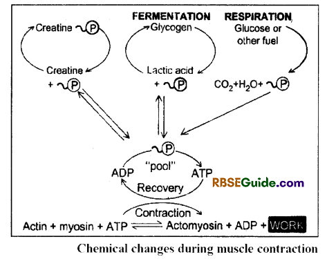
Energy Sources for Contraction
Both muscle contraction & relaxation require energy and the main source of energy is ATP. The muscles have less number of stored ATP. Hence, during contraction.ATP is obtained from other sources.
The creatine-phosphate found in the muscles provides energy instantly because it has one high energy bond. It is also called as phosphagen. This process is catalysed by an enzyme creatine kinase.
The energy required in muscle contraction is obtained by oxidation of glucose but due to deficiency of Q, this source fails to provide energy’ for long period. Hence, the fermentation of the glycogen is the long-lasting source of the energy for muscle contraction.
Glycogen fermentation results in the formation of lactic acid. This lactic acid is transported into the liver by the blood. In the liver, 80% lactic acid is recovered into the glycogen (Glyconeogenesis) and the remaining 20% lactic acid is oxidized into CO, & If, in which energy is released. This energy is used in the glyconeogenesis.
Diseases of Bones
Arthritis
Arthritis is normally joints pain, which is due to inflammation of the joints. It is a degenerative disease and occurs with age. Joints become stiff and are painful on movement. Arthrities is of three kinds –
Rheumatoid arthritis
It involves inflammation of the synovial membrane. This disease may occur be at any age. Temperature also increases at joints. Membrane secretes abnormal granules i.e. pannus. Deposition of pannus makes the joint immovable. The joints become stiff’ almost immovable due to formation of hard tissue over the cartilage.
It is a kind of autoimmune disease, in which immune system of body attack on the tissue of itself. This disease may be diagnosed by the presence of immunoglobulin, Ig M and increased erythrocyte sedimentation rate (E S R). Rheumatoid arthritis is generally in old age and affects the hands and legs of a person. For therapy, pain killer medicines are given and physiotherapy is suggestive.
Osteoarthritis
It also a disease of old age. The cartilage present at the joint is ossified, losing its flexibility. The movement of joints becomes extremely painful. Effect of osteoarthritis is more at pelvic joints, knee joints etc.
Joint bones become thin and weak & gap between ]oints is reduced. New bone formation is the main symptoms of disease due to narrowing of joint movement in the joint is reduced and becomes painful. For treatment, pain killers are given. Joints are also replaced by metallic or plastic implants in serious condition.
Gouty Arthritis
It is also a disease of joints and caused by deposition of uric acid crystals in the cavity of joints. In such patients, most of the nitrogenous wastes are excreted as uric acid rather than urea. It also makes the joints painful. Patients are advised to avoid high protein diet.
![]()
Osteoporosis
Main characteristics of the disease is the loss of bone mass means deficiency of organic part (matrix) and minerals (calcium) in the bones. The bone becomes thin, weak and loses elasticity and strength. Chances of bone fracture are increased and slight jark or impact may lead to bone fracture. It affects the whole body skeleton but pelvis, wrist, vertebrae etc are most adversely affected parts.
It increases with the age but deficiency of estrogen also caused osteoporosis in old women. It may also be caused by hormones like calcitonin, PTH and glucocorticoids.